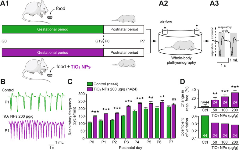
Fig. 2.
Breathing frequency is abnormally elevated in neonates that have been prenatally exposed to TiO2 NPs. A1 Schematics of protocols for non-exposed (green) and TiO2 NP-exposed (purple) pregnant mice during the gestational (G) period, and A2 subsequent whole-body plethysmography experiments performed on offspring during the first postnatal (P) week. A3 Expanded trace of the plethysmograph box pressure signal derived from a control animal. B Whole-body plethysmographic recordings of non-exposed (green), TiO2 NP-exposed (purple) neonates obtained at P1. C Bar charts showing the variation in spontaneous respiratory frequency (mean ± SEM) in non-exposed (green bars) and prenatally TiO2 NP-exposed (200 µg/g; purple bars) animals during the first postnatal week. D Bar charts illustrating the dose-dependent effect of gestational exposure to TiO2 NPs on respiratory frequency (top) and mean coefficients of variation under the four experimental conditions (bottom). Data were pooled from P0 to P6. The number of animals is indicated in each bar. **p < 0.01; ***p < 0.001. The mouse image is from Servier Medical Art website (smart.servier.com)

