Fig. 4.
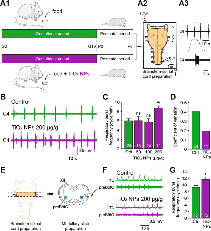
Effects of chronic maternal exposure to TiO2 NPs during gestation on respiratory-related motor burst activity in offspring ex vivo. A1 Schematics of protocols for non-exposed (green) and TiO2 NP-exposed (purple) pregnant mice during the gestational (G) period, and subsequent electrophysiological experiments performed on isolated brainstem-spinal cord preparations (A2) from offspring during the three first postnatal (P) days. A3 Raw spontaneous inspiratory-related burst activity recorded from a cervical (C4) spinal ventral root. B Raw extracellular recordings of spontaneous burst activity in a C4 ventral root of isolated preparations from unexposed (green) and prenatally TiO2 NP (200 µg/g)-exposed (purple) neonates obtained at P0. C Bar charts showing the variation in spontaneous burst frequency (mean ± SEM) in control (Ctrl, green bar) and prenatally TiO2 NP-exposed (purple bars) groups. Data were pooled from P0 to P3. The number of animals is indicated in each bar. D Mean coefficients of variation in cycle durations for the control and TiO2 NP (200 µg/g)-exposed groups. E Schematics of experimental procedures used to obtain a medullary slice preparation. F Integrated (int.) and raw extracellular burst activity recorded directly from the pre-Bötzinger complex (preBötC) in such slice preparations from unexposed (green) and prenatally TiO2 NP (200 µg/g)-exposed (purple) neonates. G Bar charts showing difference in spontaneous respiratory burst frequency (mean ± SEM) between control (Ctrl, green bar) and prenatally TiO2 NP-exposed groups (purple bar). *p < 0.05; aCSF, artificial cerebrospinal fluid; V, trigeminal nerves; IX, glossopharyngeal nerves; X, vagal nerves; XI, accessory nerves; XII, hypoglossal nuclei. The mouse image is from Servier Medical Art website (smart.servier.com)

