Fig. 1.
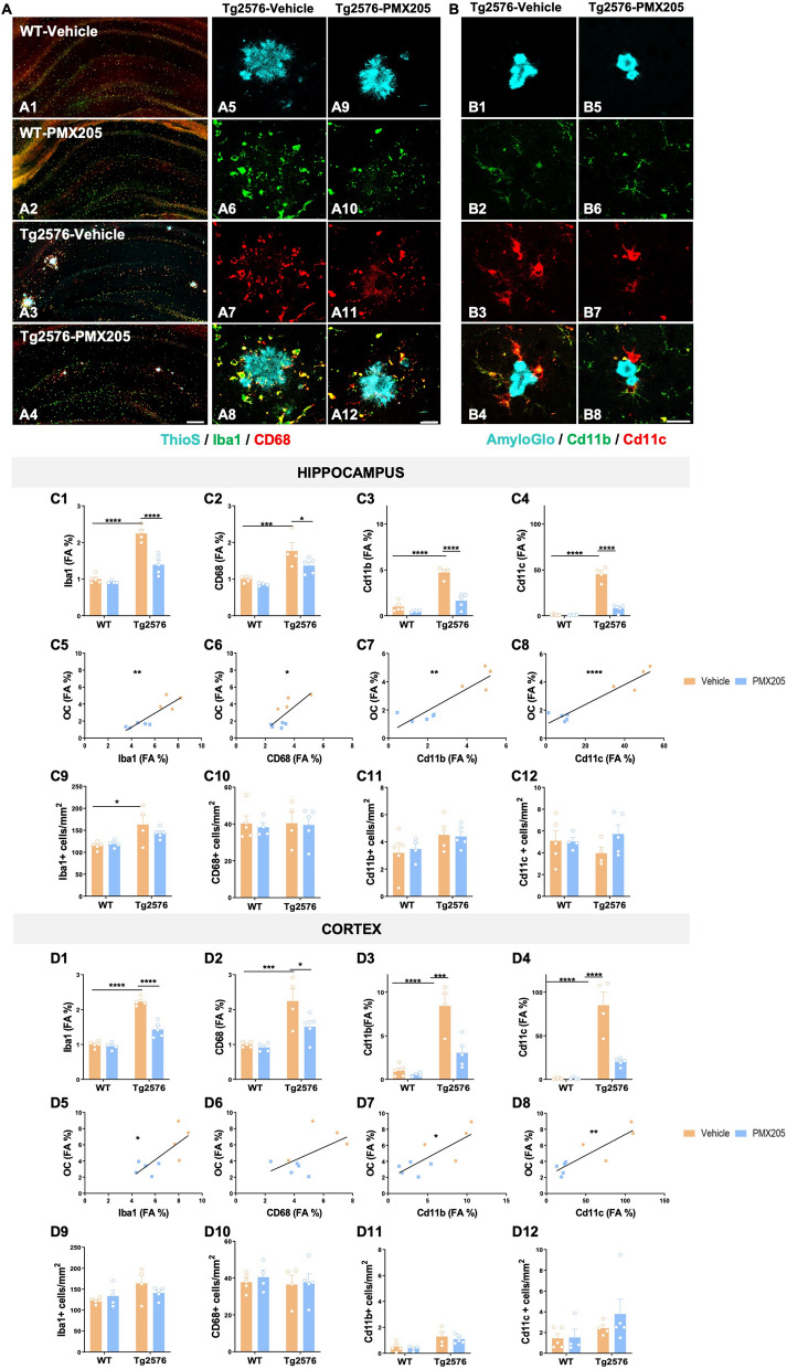
Treatment with PMX205 reduced the expression of several microglial activation markers. A–B Representative images of the hippocampus of WT-vehicle (A1), WT-PMX205 (A2), Tg2576-vehicle (A3) and Tg2576-PMX205 (A4) stained with ThioS (cyan), Iba1 (green) and CD68 (red). Inserts A5-A12 shows representative higher magnification pictures of microglial cells surrounding amyloid plaques in the hippocampus of Tg2576 vehicle/PMX205. B1–B8 Representative images of Cd11b (green) and Cd11c (red) positive microglial cells surrounding Aß deposits in the hippocampus of Tg2576-vehicle (B1–B4) or Tg2576-PMX205 (B5-B8). C–D Field Area (%) quantification of microglial markers in the hippocampus (C1–C4) and cortex (D1-D4) of Tg2576 treated with PMX205 when compared to the control group. Microglia showed a positive correlation with amyloid plaques in both, the hippocampus (C5–C8) and cortex (D5–D8) of Tg2576 mice. Quantitative analysis of microglial density (C9–C12; D9–D12) measured as number of Iba1, CD68, Cd11b or Cd11c + cells per mm2 in the hippocampus and cortex. Data are shown as Mean ± SEM and normalized to control group (WT-vehicle). Statistical analysis used a 2-way ANOVA and Pearson’s correlation test. Significance is indicated as *p < 0.05; **p < 0.01; ***p < 0.001 and ****p < 0.0001. Pearson’s R2 = 0.78 (C5); 0.58 (D5); 0.54 (C6); 0.32 (D6); 0.79 (C7); 0.61 (D7); 0.90 (C8) and 0.76 (D8). 3 sections/mouse and n = 4–5 mice per group. Scale bar: A1–A4: 200 µm; A5–A6 and B1–B2: 20 µm

