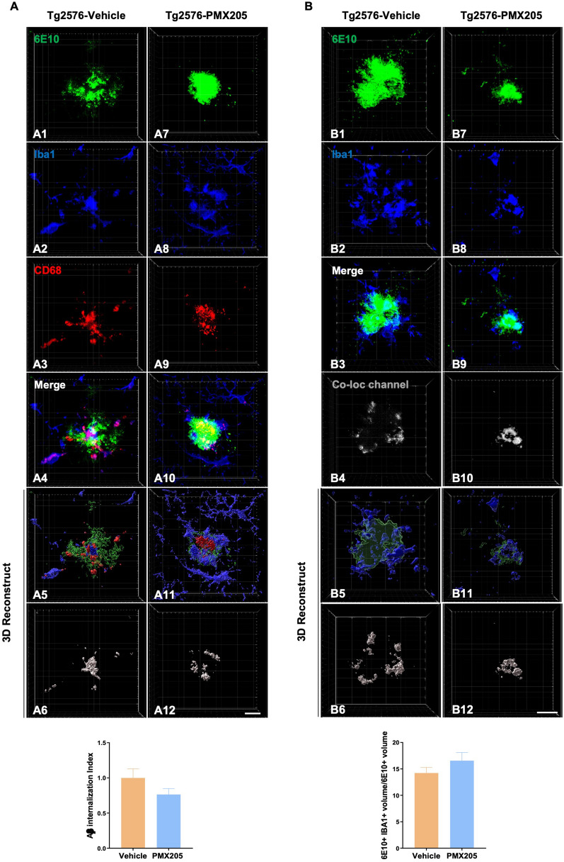
Fig. 2.
Impact of PMX205 on microglia-plaque interaction in the Tg2576 mouse model of Alzheimer’s disease. A Representative immunohistochemical images of amyloid plaques 6E10: green), microglial cells (Iba1: blue) and microglial lysosomes (CD68: red) in the Tg2576-vehicle (A1-A6) and Tg2576-PMX205 (A7-A12). Imaris 3D-reconstruction images showed a trend towards reduced internalization of Amyloid-ß by microglial cells (A6 and A12) in the Tg2576-PMX205 mice when compared to the vehicle treated group. B Representative images of amyloid plaques (6E10: green) and microglial cells (Iba1: blue) in the Tg2576-vehicle (B1-B4) and Tg2576-PMX205 (B7-B10). Tridimensional reconstruction images (B5-B6 and B11-B12) and quantification showed no difference in the engagement of microglial cells around amyloid plaques between the PMX205 treated group and the controls. Data are shown as Mean ± SEM and normalized to control group (Tg2576-PMX205). Statistical analysis used a two-tailed t-test. 15 amyloid plaques/mouse and n = 4–5 per group. Scale bar: 10 µm

