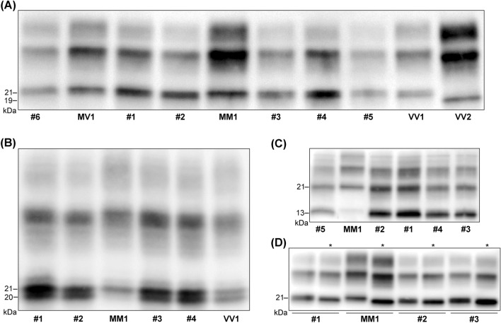
Fig. 3.
Immunoblot profiles of PK-resistant PrPSc fragments (PrPres) in the reported sCJD cases. A Immunoblot analysis of PK-treated frontal cortex homogenates by standard SDS-PAGE gel electrophoresis (running gel of 6.5 cm). The six reported cases (#1–6) are compared with sCJD cases of the MM1, MV1, VV1, and VV2 groups. Note the slightly faster migration of unglycosylated PrPres in cases 1–6 and the VV1 compared to the MM1 and MV1. B Immunoblot analysis of PK-treated frontal cortex homogenates by high-resolution SDS-PAGE gel electrophoresis (running gel of 15 cm). A sCJD MM1 and a sCJD VV1 are included for comparison with cases #1–4. The higher resolution of the gel shows that unglycosylated PrPres in VM1 and VV1 cases comprises a doublet (i.e., two bands of 21 and 20 kDa), explaining the slightly faster migration compared to MM1 and MV1 cases (they show one band of 21 kDa) C Immunoblot profile of PrPres comprising PrPSc type 1 and the 12–13 kDa C-terminal fragments. Note the higher proportion (i.e., relative amount) of C-terminal fragments in VM1 and VV1 cases compared to MM1 and MV1 (see Table 3 for the quantitative data). D Comparison of the electrophoretic mobility of PK-resistant PrPSc after digestion at pH 6.9 or 8.0 (see * labels). There is a more consistent shift in migration (i.e., faster) when PK digestion is performed at pH 8 in MM1 and MV1 cases compared to VM1 and VV1. The immunoblots shown in A, B, and C are labeled by the N-terminal mAb 3F4. In contrast, the immunoblot in panel C is stained with the C-terminal antiserum 2301. Approximate molecular masses are in kilodaltons

