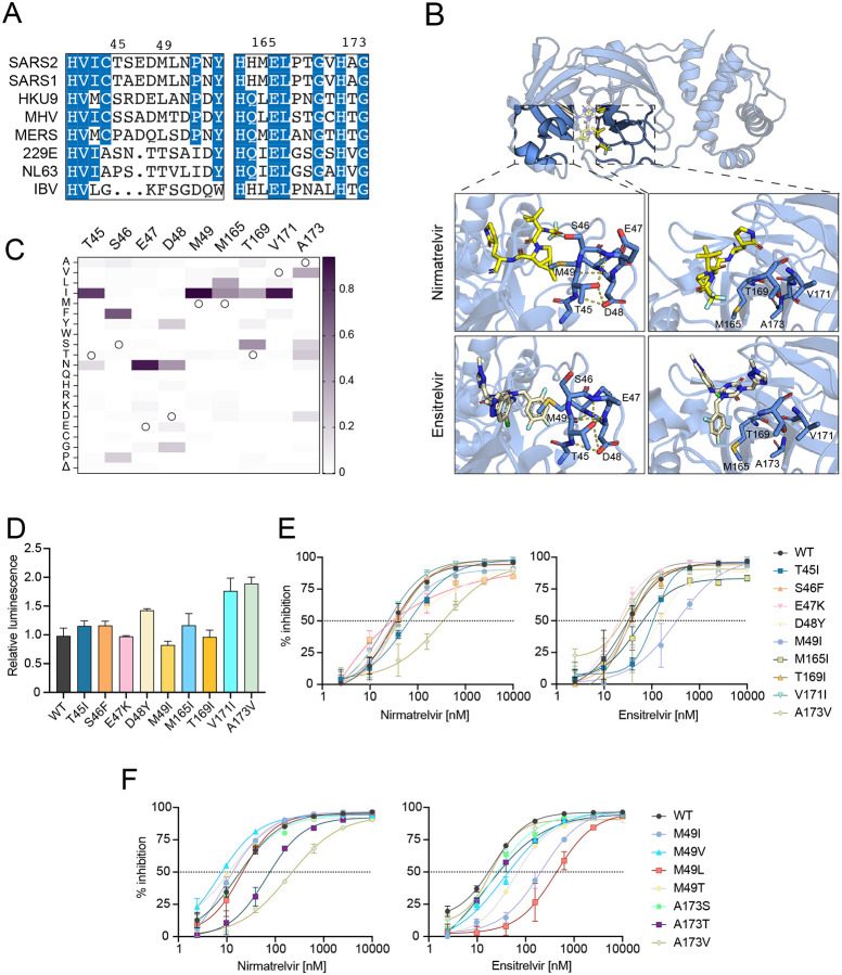
Fig. 2. Variable active site residues elicit differential resistance to nirmatrelvir and ensitrelvir.
(A) Alignment of coronavirus Mpro amino acid sequences spanning residues 41–54 and 163–174 (based on SARS2 Mpro residue position).
(B) Structure of SARS2 Mpro and inhibitors highlighting variable residues that can be mutated (PDB: 7SI9 and 7VU6 for nirmatrelvir and ensitrelvir, respectively).
(C) Relative frequency of amino acid changes at tested variable residues, excluding the respective WT residue (open circle), in SARS2 genomes (1-July-2022, GISAID database).
(D) Relative luminescence of cells expressing Src-Mpro-Tat-fLuc variants in the absence of inhibitor.
(E-F) Dose-response curves of variants using the live cell Src-Mpro-Tat-fLuc assay with 4-fold serial dilution of inhibitor beginning at 10 μM (data are mean +/− SD of biologically independent triplicate experiments).

