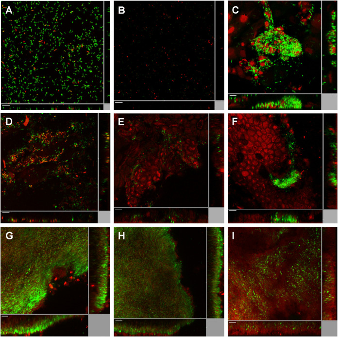
FIG 2.
Induction of the c-di-GMP reporter fusion pcdrA-gfp in P. aeruginosa during mouse corneal infection. (A) Planktonic PAO1/plac-gfp cells; (B) planktonic PAO1/pcdrA-gfp cells; (C) PAO1/plac-gfp cells at 8 h postinfection (8 hpi) of mouse cornea; (D) PAO1/pcdrA-gfp cells at 2 hpi of mouse cornea; (E) PAO1/pcdrA-gfp cells at 4 hpi of mouse cornea; (F) PAO1/pcdrA-gfp cells at 8 hpi of mouse cornea; (G) PAO1/pcdrA-gfp cells at 1 dpi of mouse cornea; (H) PAO1/pcdrA-gfp cells at 4 dpi of mouse cornea; (I) PAO1/pcdrA-gfp cells at 7 dpi of mouse cornea. SYTO 62 was used to stain host cells as well as P. aeruginosa cells lacking fluorescence. Green fluorescence represents constitutive expression of plac-gfp (A and C) and expression of the pcdrA-gfp reporter fusion (B and D to I), and red fluorescence represents SYTO 62 staining. Experiments were performed in triplicate, and a representative image for each condition is shown. Scale bars, 10 μm.

