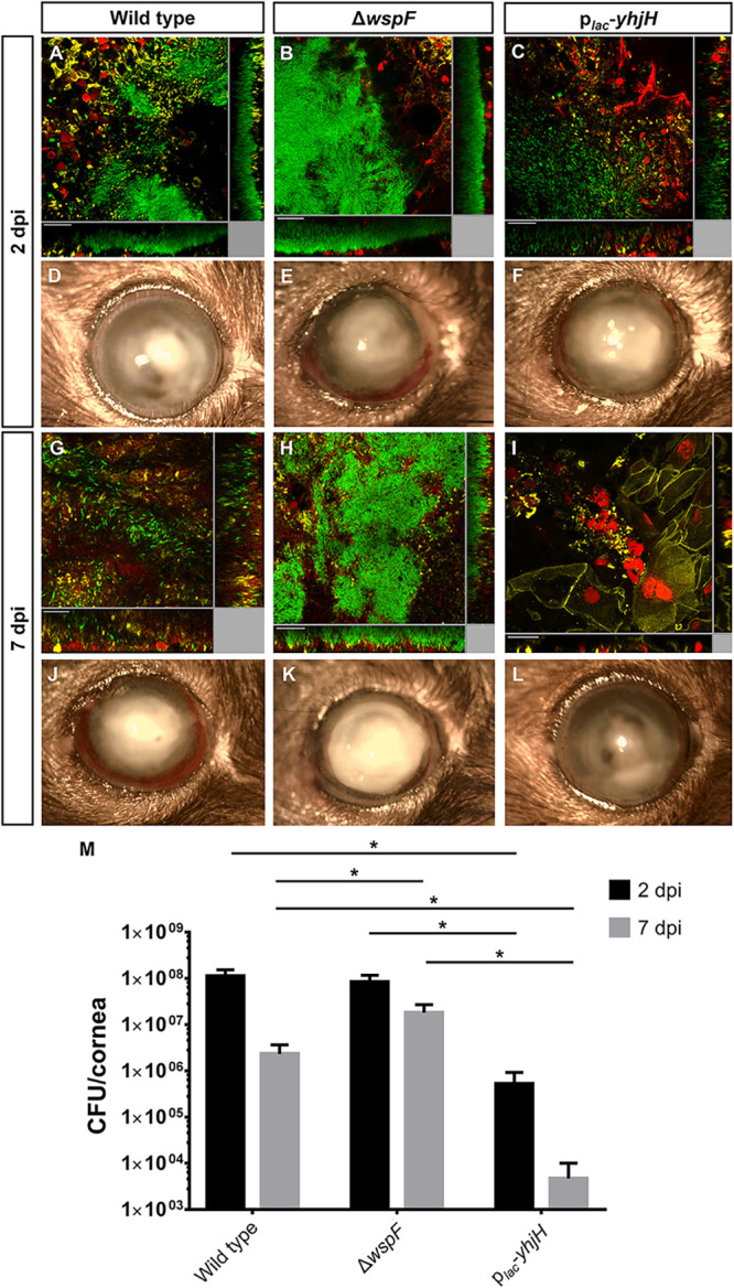
FIG 3.

Bacterial load of P. aeruginosa in mouse cornea after infection with the wild-type PAO1, ΔwspF mutant, and plac-yhjH strains at 2 dpi and 7 dpi. (A to C and G to I) Confocal images of infected mouse cornea with the PAO1 wild-type, ΔwspF, and plac-yhjH strains at 2 dpi and 7 dpi. The P. aeruginosa bacteria were tagged with plac-gfp in a mini-Tn7 construct. The red fluorescence represents the staining of lysosomes by LysoTracker Red DND-99. The yellow fluorescence represents Alexa Fluor 635-phalloidin, which stains F-actin of the eukaryotic cells. (D to F and J to L) Slit lamp images of infected mouse cornea with the wild-type PAO1, ΔwspF, and plac-yhjH strains at 2 dpi and 7 dpi. Experiments were performed in triplicate, and a representative image of each condition is shown. Scale bars, 20 μm. (M) CFU of corneas infected with the wild-type PAO1, ΔwspF, and plac-yhjH strains at 2 dpi and 7 dpi. Mean values and SD from triplicate experiments are shown. *, P < 0.01, Student's t test.
