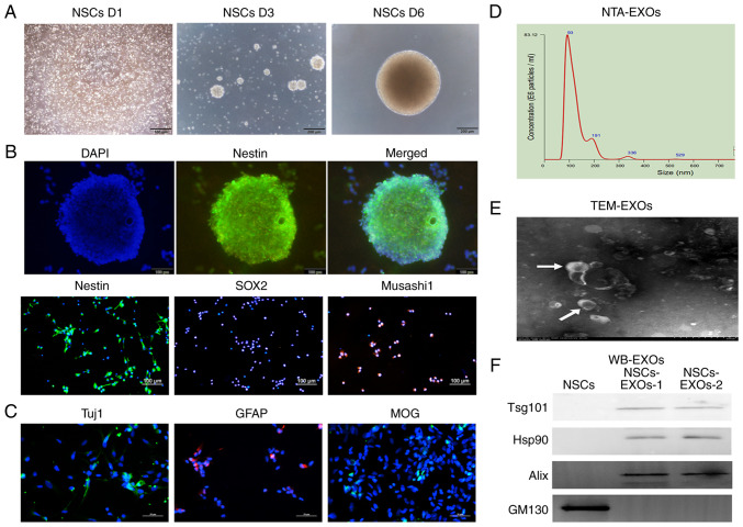
Figure 1.
Characterization of exosomes derived from NSCs. (A) The morphology of NSCs was recorded on day 1, 3 and 6 after cell thawing. Scale bar, 200 µm. (B) Immunofluorescence detected the stem cell markers, Nestin, SOX2 and Musashi1. Scale bar, 100 µm. (C) Immunofluorescence detected neurons (Tuj1), astrocytes (GFAP) and oligodendrocytes (MOG). Scale bar, 50 µm. (D) NTA analysis revealed the particle distribution of exosomes derived from NSCs. (E) The exosomes was detected using TEM. Scale bar, 100 nm. (F) Western blot analysis of the exosomal markers, Tsg101, Hsp90 and Alix, and the Golgi marker, GM130. NSCs, neural stem cells; NTA, nanoparticle tracking analysis; MOG, myelin oligodendrocyte glycoprotein; GFAP, glial fibrillary acidic protein; TEM, transmission electron microscopy; EXOs, exosomes.

