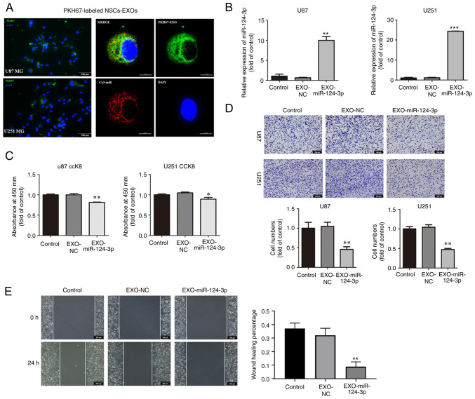
Figure 2.
NSC-EXOs loaded with miR-124-3p suppress glioma cell proliferation, invasion and migration. (A) EXOs were isolated from neural stem cell supernatant, dyed with PKH67 (green), and co-cultured with glioma cells for 24 h. Subsequently, they were dyed with DAPI (blue) and examined (EXOs labeled with PKH67, miR labeled with Cy5; original magnifications, ×100 (left panel) and ×400 (right panel). (B) miR-124-3p expression was detected using reverse transcription-quantitative PCR following incubation of the glioma cells for 48 h. (C) The proliferative ability of U87 and U251MG cells was tested using CCK-8 assay. (D) Transwell invasion assays in glioma cells were used to determine cell invasion. (E) Cell migration was detected through a wound-healing assay in glioma cells (original magnifications, ×100) Data represent the mean ± SD from three independent experiments. *P<0.05, **P<0.01 and ***P<0.001, statistically significant differences between the NC and EXO-miR-124-3p group. NC, miR negative control; NSC, neural stem cell; EXOs, exosomes.

