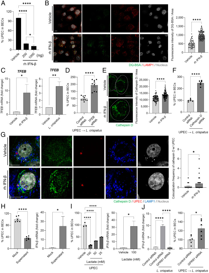
Fig. 2.
Intracellular bactericidal action of type I IFN is mediated by lysosomal maturation. (A) Intracellular UPEC killing by type I IFN. Human 5637 BECs were treated with L. crispatus at various MOI (200). After 6 h of infection, the bacterial burden was measured 20 h after treatment with recombinant human (rh) IFN-β (200 or 1000 ng). (B) Lysosomal maturation by IFN-β. Human 5637 BECs were treated with rh IFN-β (500 ng). After 24 h of treatment, cells were exposed to DQ-BSA (green) for 6 h and then immunostained using an anti-LAMP1 (red) antibody for confocal microscopic imaging. (B, Right) The mean fluorescence intensity of DQ-BSA was measured in random fields. (C) After treatment with rh IFN-β (500 ng) for 6 h, the expression of TFEB mRNA in cells treated with IFN-β or L. crispatus was compared to that in vehicle-treated control using qPCR. (D) TFEB mediated L. crispatus-induced intracellular UPEC killing in BECs. After human BECs were transfected with control or TFEB siRNA, UPEC-infected BECs were exposed to L. crispatus. The intracellular number of UPEC in treated BECs was assessed after 16 h. (E) Enhanced cathepsin D expression was assessed by treating BECs with rh IFN-β (500 ng) for 24 h followed by anti-cathepsin D (green) immunostaining for confocal imaging. The dotted line indicates the periphery of BECs. (E, Right) The fluorescence intensity of cathepsin D was measured in random fields. (F) Cathepsin D regulates the size of intracellular UPEC population. After human 5637 BECs were transfected with control or cathepsin D siRNA, UPEC-infected BECs were exposed to L. crispatus. The intracellular number of UPEC in treated BECs was assessed after 16 h. (G) UPEC-infected BECs were treated for 12 h with IFN-β (500 ng) and then immunostained for cathepsin D (green), UPEC (red), and LAMP1 (blue). (G, Right) The frequency of colocalization between cathepsin D and UPEC was measured in random fields. (H) Effects of CFS of L. crispatus on human BECs. (H, Left) UPEC-infected human BECs were CFS or mock treated for 16 h, and the bacterial load was determined. (H, Right) The relative change of IFN-β mRNA expression by human BECs was analyzed using qPCR after 6 h of exposure to CFS of L. crispatus. (I) Effects of lactate on human BECs. (I, Left) UPEC-infected human BECs were treated with lactate in a dose-dependent manner or mock treated for 16 h; thereafter, the intracellular bacterial load was determined. (I, Right) The relative change in expression levels of IFN-β mRNA in human BECs was assessed using qPCR after 6 h of treatment with lactate (100 mM). (J) The lactate effect on BECs is mediated by GPR81. After human 5637 BECs were transfected with control or GPR81 siRNA, (J, Left) the relative change in IFN-β mRNA expression levels was assessed using qPCR after 6 h of treatment with L. crispatus. (J, Right) UPEC-infected BECs were exposed to L. crispatus (MOI 1000), and the intracellular number of UPECs in treated BECs was assessed after 16 h. Error bars in each bar graph show mean ± SEM. Each experiment (A–J) was independently repeated two to three times with similar results. Data were analyzed by unpaired two-tailed Student t test (B–J) or an ordinary one-way ANOVA (A and I) with Tukey’s multiple comparison posttest. *P < .05; **P < .01; ****P < .0001. Scale bars: 10 μm.

