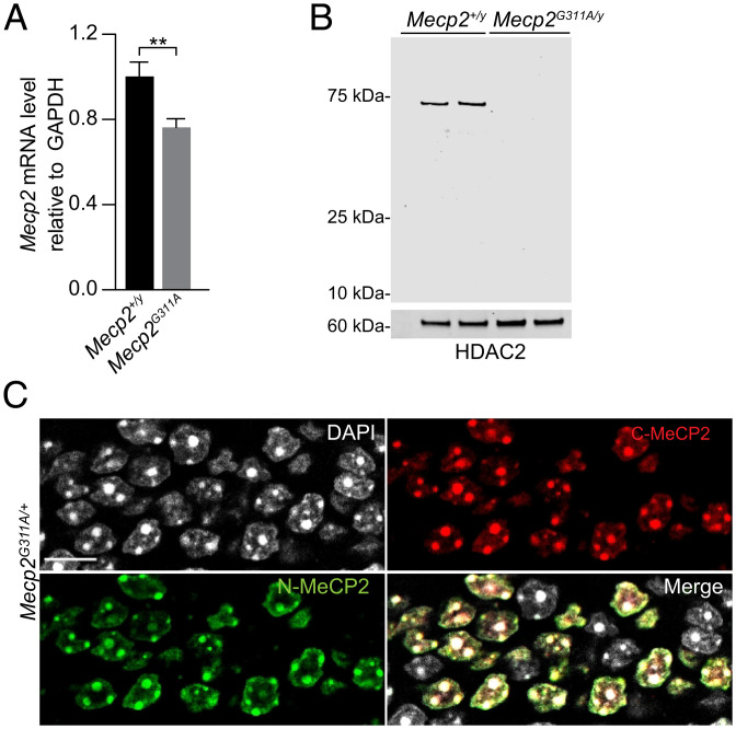
Fig. 1.
Analysis of Mecp2 RNA and protein in the mutant Mecp2G311A mouse model. (A) Real-time qRT-PCR of Mecp2 RNA in indicated genotypes. n = 3 mice per genotype. Histogram values are mean ± SD, **P < 0.01 using Student’s t test. (B) Western blot of nuclear lysates prepared from brains of postnatal day 56 male mice. n = 2 mice per genotype. The blot was probed with antibodies directed against the N terminus of MeCP2 and HDAC2 (for the loading control). (C) Confocal images acquired from a dentate gryus section of a female Rett syndrome mouse showing mosaic staining for MeCP2 detected by immunolabeling for the N and C termini of MeCP2. (Scale bar, 10 µm.)

