Fig. 4.
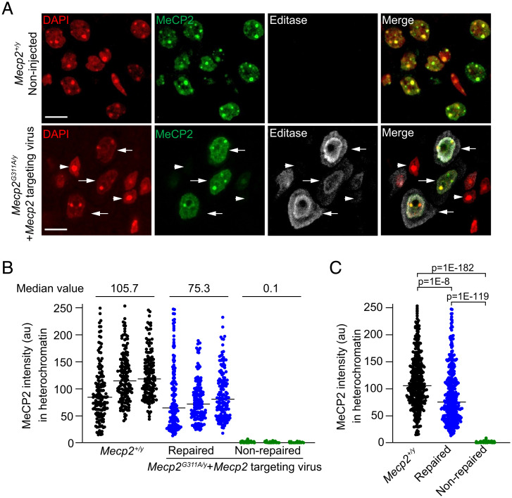
Mecp2-targeted RNA editing rescues MeCP2 protein expression and function in the brainstem. (A) Confocal images of nuclei in brainstem sections from noninjected wild-type Mecp2+/y male mice (Upper) and mutant Mecp2G311A/y mice injected with the Mecp2-targeting virus (Lower). DAPI staining defines nuclear compartment and heterochromatic foci. Arrows indicate repaired cells expressing MeCP2 and Editasewt; arrowheads indicate nonrepaired cells lacking both MeCP2 and Editasewt expression. Editasewt intensities appear greater near the nuclear membrane and in the cytoplasm. (Scale bar, 10 µm.) (B) Quantification of immuno-labeled MeCP2 associated with heterochromatic puncta for three individual wild-type mice and three mutant mice injected with the Mecp2-targeting virus. Each dot represents the mean of MeCP2 intensities within all heterochromatic foci in an individual nucleus. Median values are indicated for the comparisons; n = 150 cells per mouse. Note that the repaired and nonrepaired nuclei are in the same section of an injected mutant mouse. The criterion for a nonrepaired nucleus is based on estimates from sections from noninjected mutant mice. (C) Histogram showing pooled results for the three mice associated with each condition (n = 450 cells per condition). Horizontal lines represent the median intensities. Statistical comparisons of distributions are by Kruskal–Wallis and Dunn’s multiple comparisons tests with a Bonferroni correction.

