Fig. 5.
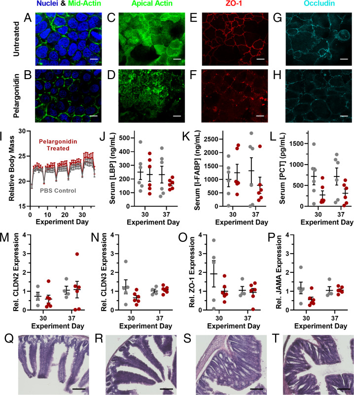
Pelargonidin induces reversible, nontoxic opening of intestinal tight junctions. (A) Compared to untreated Caco-2 cells, (B) pelargonidin-treated monolayers showed no difference in morphology for nuclei or midcell actin. (C and D) However, actin at the apical surface, as well as the tight junction proteins (E and F) ZO-1 and (G and H) occludin rearranged into more punctate forms as a result of pelargonidin treatment. (I) During 1 mo of daily pelargonidin treatment, mice did not lose weight compared to control animals. Periodic weight loss was observed in both control and treated groups as a result of overnight fasting for weekly checkups. (J) Treated mice did not develop elevated levels of the inflammation markers LBP, (K) I-FABP, or (L) PCT. qRT-PCR revealed no statistical difference in mRNA expression of the tight junction proteins (M) Claudin 2, (N) Claudin 3, (O) ZO-1, or (P) JAMA in the small intestines of control and pelargonidin-treated mice. (Q) Representative histological images of duodenal sections of the small intestines from control mice and (R) pelargonidin-treated mice displayed no tissue damage resulting from treatment. (S) There were also no discernable histological differences between proximal colon samples from control and (T) treated mice. (White scale bars, 10 µm; black scale bars, 100 µm.) Error bars represent SEM (n = 4 to 12). For I–P, no comparisons between treated and control mice achieved statistical significance by two-tailed t test with Welch’s correction.

