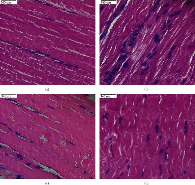
Figure 11.

(a–d). Histological sections of the gastrocnemius muscle in (a) control rats reveal normal parallel acidophilic myofibers grouped in bundles; (b) diet-induced diabetic rats show crumbling myofibers with many peripheral flattened nuclei; (c) widespread myofibers with peripheral nuclei in diet-fed diabetic rats treated with β-sitosterol; (d) aligned myofibers with few crushed marginal nuclei in diet-induced diabetic rats treated with metformin.
