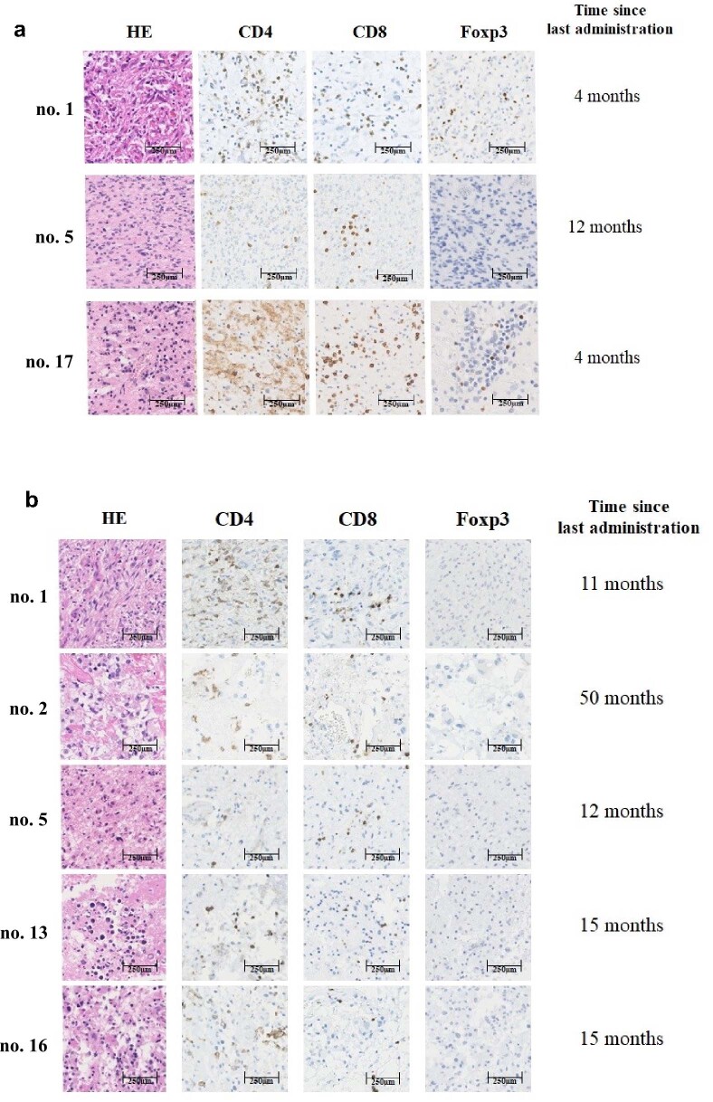
Extended Data Fig. 8. Histology of regrown tumors at reoperation and brain lesions at autopsy.
a After G47∆ therapy, 3 patients underwent craniotomy for tumor resection of regrown tumors (no. 1 and no. 17 at 4 months and no. 5 at 12 months after the last G47∆ administration). Infiltrating numbers of CD4 + and CD8 + lymphocytes remained abundantly increased in the two cases at 4 months and to some extent in the case at 12 months. Increased numbers of Foxp3+ cells were found in the two cases at 4 months and both underwent reoperation, but not found in the case at 12 months. Each is representative of 3 tissue samples. b Among 5 patients who underwent autopsy, 4 patients died of tumor progression (no. 1, no. 2, no. 5 and no. 16) and an infiltration of CD4+ and CD8+ lymphocytes and a low number of Foxp3+ cells persisted at autopsy 11 to 50 months after the last G47Δ administration. In patient no. 13 whose target lesion was well controlled at the time of death, necrosis and calcification, but no viable tumor cells, were found in the brain at autopsy 15 months after the last G47∆ administration. Each is representative of 3 tissue samples.

