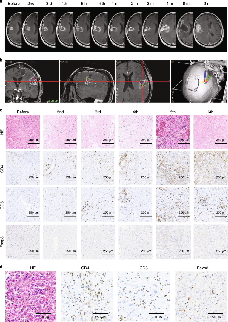
Fig. 2. Representative case (patient no. 1) treated with G47Δ.
a, MRI images at indicated observation time points. Characteristic MRI changes were observed at every G47Δ administration, that is, a clearing of contrast-enhancement at the injection site and an enlargement of the entire target lesion. G47∆ was injected to different coordinates from previous injections, so the area of contrast-enhancement clearing increased as the doses increased, leading to a large hollow within the target lesion with an increase in diameter after six G47∆ doses. The MRI changes ceased after the last G47∆ injection and the target lesion stayed stable until 4 months after G47∆ therapy, when the tumor showed a regrowth. The regrown tumor was resected at 6 months but further regrew at 9 months, and the patient died of exacerbation of the disease at 16.2 months after G47∆ initiation. b, Planning MRI using StealthStation Surgical Navigation System at the 6th dose, displaying administration routes from the 2nd to 6th dose (10 injections) overlaid in the same image. c, Histology of biopsy specimens. Biopsies were performed before indicated injections and were obtained from coordinates different from previous G47∆ injections. CD4+ and CD8+ lymphocytes infiltrating within the tumor increased abundantly in number as G47∆ injections were repeated. In contrast, the number of tumor-infiltrating Foxp3+ cells remained low throughout repeated G47∆ injections. Representative of four biopsy specimens. d, Histology of resected tumor at regrowth. The numbers of tumor-infiltrating CD4+ and CD8+ lymphocytes remained high in the regrown tumor resected 6 months after the last G47∆ administration. A higher number of Foxp3+ cells are observed in the tumor at regrowth than in tumors during G47∆ treatment. Representative of three tissue samples. HE, haematoxylin and eosin; m, month(s).

