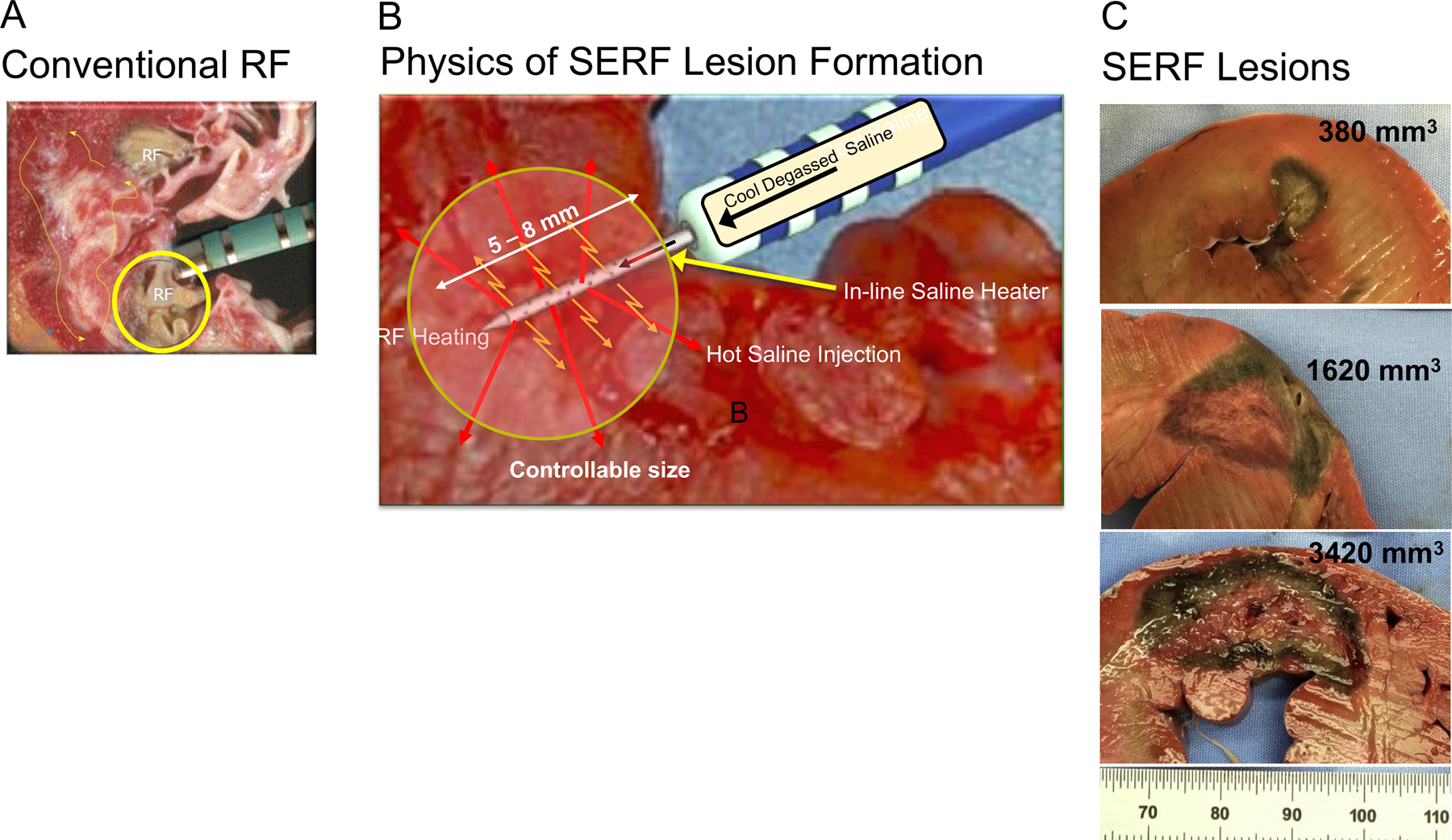
Figure 2.

Lesion Creation with Standard RF and SERF Heated Saline Ablations. Panel A shows RF catheter tip/tissue contact and a representative endocardial surface lesion using a standard catheter design. Panel B is a rendition showing the positioning of the SERF catheter along with the needle extension into the myocardium. Heated saline is injected through side holes into the tissue, markedly increasing convective heating (orange arrows) during RF application to the needle. Panel C shows a representative spectrum of needle ablation lesions with size and depth of extension to the mid and epicardial myocardium from a previous animal model. [31, 32] [29] [33] [28–30]
