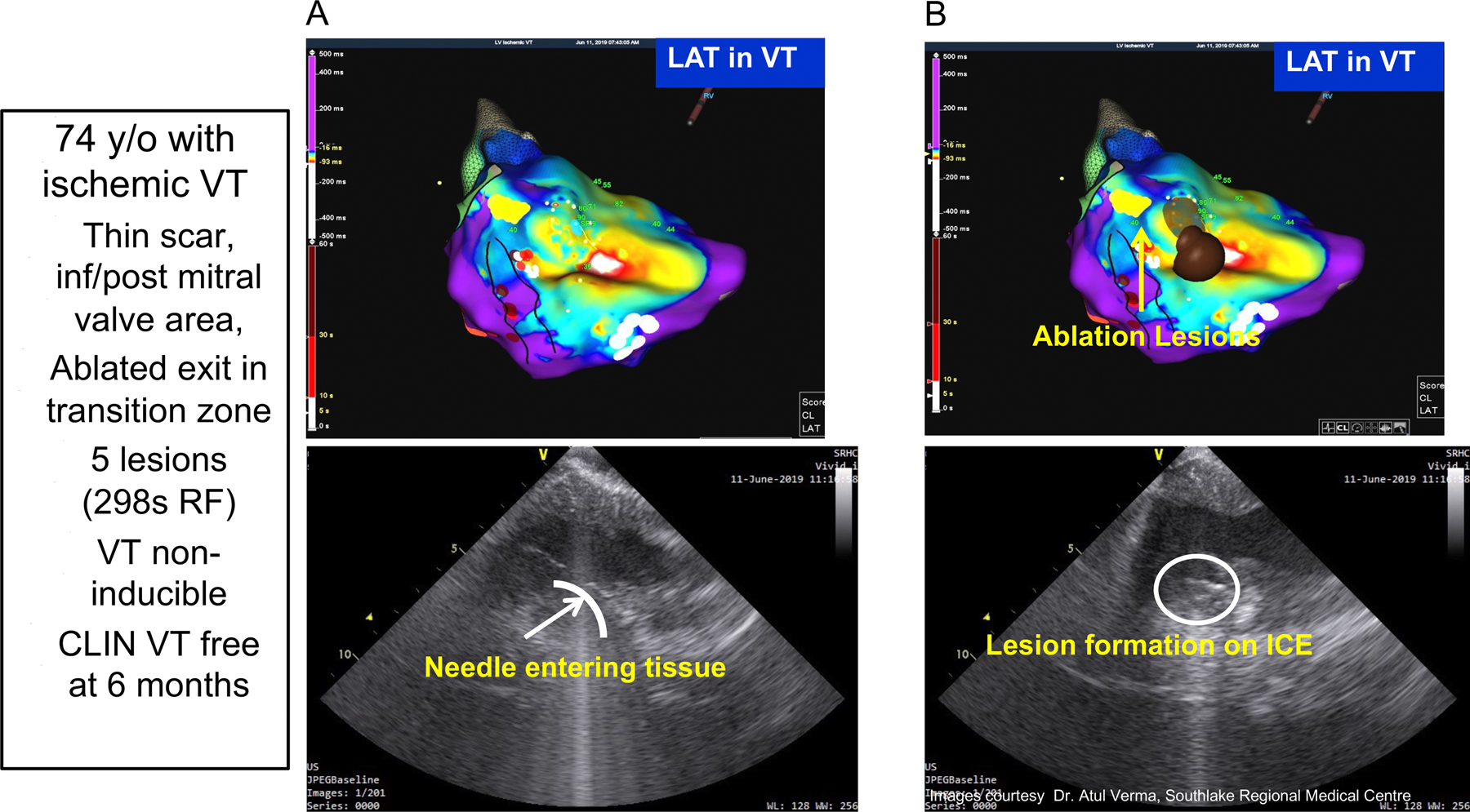
Figure 4.

Mapping and Intracardiac Ultrasound Imaging from a patient with prior infero-posterior myocardial infarction and recurrent VT that had failed to be controlled by prior ablation procedures. Panel A shows the activation map of the targeted VT. Panel B shows the site of ablative lesions. Panel C shows a pre-ablation ICE image of the catheter tip/issue positioning and the inserted needle into the underlying myocardium. Panel D show the evolving ablation lesion indicated by the increasing contrast and intensity of the local tissue seen on ICE. Five needle ablation lesions were applied at the endocardial break out region and abolished this inducible VT, which remained absent during 6 month follow-up.
