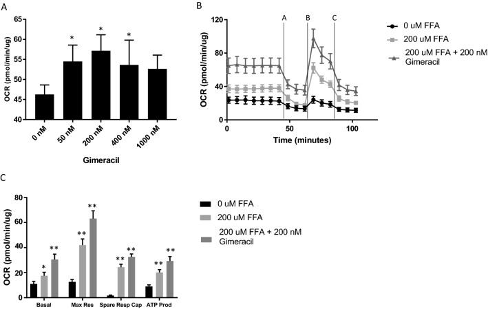
Figure 3.
Extracellular flux analysis demonstrates enhanced mitochondrial functionality in PHHs treated with Gimeracil. Mitochondrial stress test involves the addition of oligomycin A (2.0 µM, time interval A) FCCP (2.0 µM, time interval B) and Rotenone Antimycin A (0.5 µM, time interval C). * indicates adjusted p-value < 0.05 and ** indicates adjusted p-value < 0.0001. (A) PHHs treated with increasing concentrations of the Gimeracil in the presence of 200 µM palmitic and oleic acid demonstrate a significant increase in oxygen consumption rate (OCR) at 50 nM (adjusted p-value 0.0173), 200 nM (adjusted p-value 0.0017) and 400 nM (adjusted p-value 0.0754). The increased observed at 1000 nM is not statistically significant (adjusted p-value 0.0754). (B) OCR normalized to total cellular protein demonstrates FFA treatment (light gray square) increased OCR and 200 nM Gimeracil (dark gray triangle) further increased OCRrelative to no treatment (black circle). (C) Basal oxygen consumption increased with both FFA treatment (light gray, adjusted p-value 0.0064) and FFA and Gimeracil treatment (dark gray, adjusted p-value < 0.0001). Maximal respiration, spare respiratory capacity and ATP production increased with FFA treatment (adjusted p-value < 0.0001 for all three metrics) and increased further with Gimeracil treatment (adjusted p-value < 0.0001 for all three metrics).

