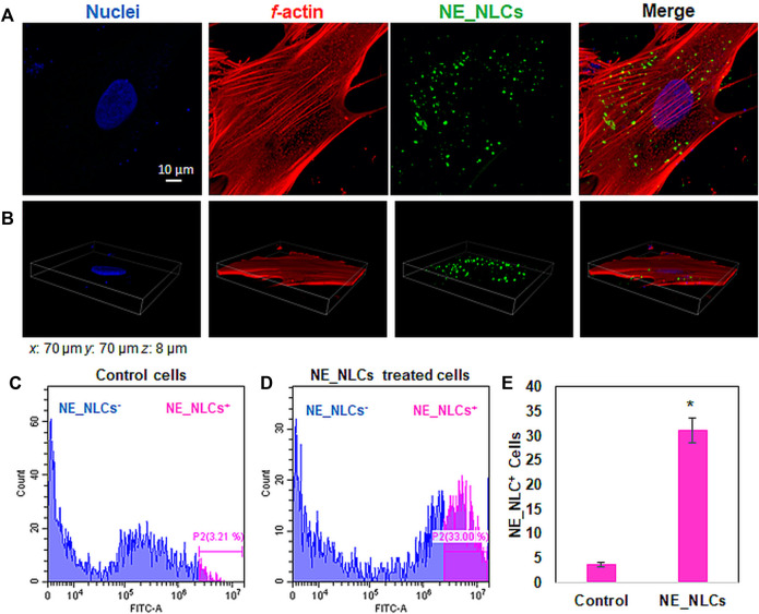
FIGURE 4.
Representative confocal images of HDFs treated for 24 h with NE_NLCs 100 μg/ml (NE_NLCs in green, f-actin in red, and nuclei in blue): single z-stack (A) and 3D rendering (B) images. Representative flow cytometry results of control (C) and NE_NLCs-treated (D) HDFs, as well as quantitative analysis (E). Data are represented as mean values ±standard deviation (n = 3; *p < 0.05).

