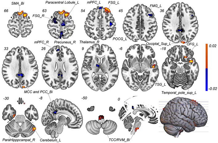
Figure 3.
Discriminative features to discriminate MWoA patients and HCs. Red means positive weight and blue means negative weight. The main weight brain areas were located in TCC/RVM, thalamus, PFC, and TSG/TMG. Bi, bilateral; FMG, frontal middle gyrus; FSG, frontal superior gyrus; L, left; MCC, middle cingulate cortex; mPFC, medial prefrontal gyrus; OFG, orbitofrontal gyrus; PCC, post cingulate cortex; POCG, postcentral gyrus; R, right; RVM, rostral ventromedial medulla; SMA, supplementary motor area; Sup, superior; TCC, trigeminal cervical complex; TMG, middle temporal gyrus; TSG, superior temporal gyrus.

