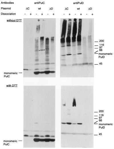
FIG. 4.
Formation of 110-kDa band upon cross-linking of cells from maltose-induced cultures of strains producing PulC protein. Whole cells of strains carrying pCHAP1229 (ΔpulC [ΔC]), pCHAP231 (wild type [wt]), or pCHAP1226 (ΔpulD [ΔD]) were incubated with 0.3 mM DSP for 15 min, extracted with phenol (dissociation), and dissolved in SDS-PAGE sample buffer without or with DTT to reduce the disulfide bond formed by the cross-linker. Proteins reacting with PulC or PulD antibodies were detected by SDS-PAGE and immunoblotting, using the same nitrocellulose sheet. The positions of PulC and PulD monomers are indicated, as are the positions of the ca. 110-kDa band that appears only after cross-linking of cells carrying pCHAP231 (dot) and molecular size markers (indicated at the right in kilodaltons).

