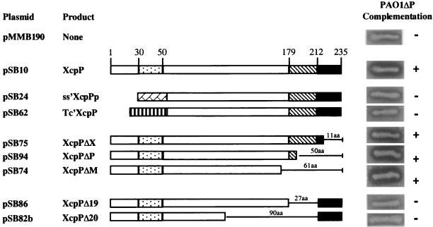
FIG. 2.
Schematic representation and characterization of the various forms of XcpP. Sizes of the deletions and amino acid (aa) residue positions are indicated. XcpP domain motifs are as in Fig. 1. Also shown are the LasB signal peptide ( ) and TetA N terminus (
 ). For each construct, complementation of PAO1ΔP is shown by halo formation on skim milk plates containing 300 μg of carbenicillin per ml and 2 mM IPTG.

