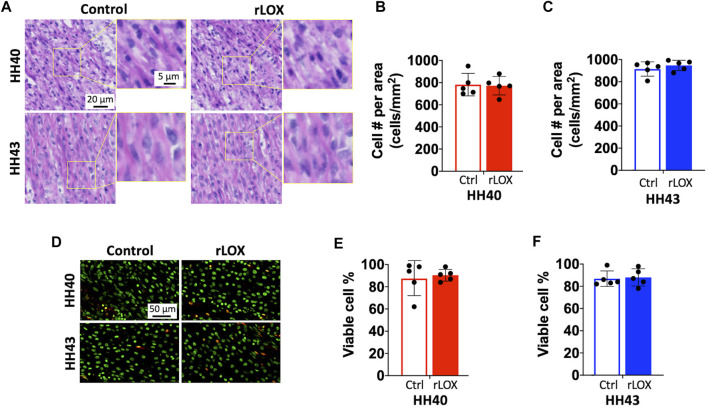
FIGURE 3.
Cell number and viability of HH40 and HH43 tendon explants were similar between rLOX and Ctrl treatments. (A) Representative images of H&E-stained HH40 and HH43 tendons after rLOX and Ctrl treatments. High magnification images showed intact cell nuclei and lack of cell debris. (B,C) Cell density was not affected by rLOX treatment in both HH40 (B) and HH43 (C) tendons. (D) Representative images of TUNEL- and Hoescht 33342-stained HH40 and HH43 tendons after rLOX and Ctrl treatments. TUNEL-positive cells were red and Hoescht 33342-positive cells were pseudo-colored green. (E,F) Percentages of viable cells in both HH40 and HH43 tendons were similar between rLOX and Ctrl treatments. Statistically significant differences were determined by Student t-test with p < 0.05. n = 5 per stage and treatment group.

