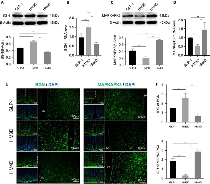
FIGURE 7.
Comparison of protein and gene expression of Bgn and Mapkapk3 in the hypothalamus. (A) WB and protein expression of Bgn in hypothalamus; (B) gene expression of Bgn in hypothalamus; (C) WB and protein expression of Mapkapk3 in hypothalamus; (D) gene expression of Mapkapk3 in hypothalamus; (E) representative figures of Bgn and Mapkapk3 in hypothalamus of rats in each group; (F) comparison of IOD of Bgn and Mapkapk3 in hypothalamus; *P < 0.05, **P < 0.01. 3V, the 3rd ventricle. WB, Western blotting; Bgn, biglycan; Mapkapk3, mitogen-activated protein kinase activated protein kinase 3; IOD, integrated optical density.

