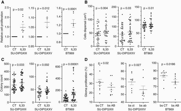
Figure 4.
IL33 impacts DIPG proliferation, growth in 3D, and colony formation. (a) Proliferation of DIPG cells over a period of 72 h with IL33 (50 ng/ml) or control condition (CT). Proliferation was measured using CellTiter-Glo luminescence. The data are presented as the fold change in proliferation with IL33 treatment compared to control, each point represents the average of 8 repeats. (Wilcoxon’s signed-rank test; SU-DIPGXIII n = 8, SU- DIPGXXV n = 6, BT869 n = 8). (b) Cells’ dispersal diameter refers to the expansion of each DIPG line in a Matrigel–-TSM mixture (1:1) with IL33 treatment, in comparison to the CT. The data were obtained by placing a concentrated droplet of DIPG cells in the center of each gel–TSM droplet and measuring the area of the DIPG droplet after 2 h (for a time zero measurement), and then comparing to the final area after 7 days. Images were obtained and the area measured, using ImageJ (Fisher’s Combined Probability Test; SU-DIPGXIII n = 6, SU-DIPGXXV n = 6, BT869 n = 5. Within each trial, a Mann–Whitney U test was performed to obtain the p-values, n = 7 droplets. One tailed). (c) DIPG colony count per well, after a treatment period of 5–7 days with IL33 in comparison to control. Quantification was performed with Incucyte® S3 Live-Cell Analysis system (Fisher’s Combined Probability Test; SU-DIPGXIII n = 11, SU-DIPGXXV n = 8, BT869 n = 6. Within each trial, a Mann–Whitney U test was performed to obtain the p-values, n = 6-8 wells. One tailed). (d) mIL33 neutralization trial in ex vivo slice cultures. Flow cytometry analysis of CellTrace dye staining of DIPG cells, injected with 1 μg/ml of mIL33 antibody or isotype control antibody. The cells were injected into two sides of the brainstem from a single P2 mouse brain, then slice cultures were prepared. After 72 h, the proliferation of DIPGs was measured (Wilcoxon’s signed-rank test, one-tailed; SU-DIPGXIII n = 10, SU- DIPGXXV n = 10, BT869 n = 11, when n represents a single brain).

