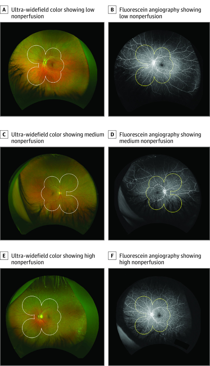
Figure 1. Examples of Eyes in the Low-, Medium-, and High-Nonperfusion Subgroups.
Representative paired ultra-widefield color and fluorescein angiography (FA) images of eyes in the low, medium, and high tertile groups of retinal nonperfusion. White border on the color and yellow border on the FA represent the Early Treatment Diabetic Retinopathy Study (ETDRS) areas border. A and B, Eye with low nonperfusion. The baseline Diabetic Retinopathy Severity Scale (DRSS) level within the ETDRS area was moderate nonproliferative diabetic retinopathy (NPDR), and there was no progression at 4 years. Predominantly peripheral lesions on FA (FA PPL) were absent. C and D, Eye with medium nonperfusion. The baseline DRSS score was moderate NPDR with progression to very severe NPDR at year 2. Color PPL and FA PPL were present. E and F, Eye with high nonperfusion. The baseline DRSS score was moderate NPDR with progression to very severe NPDR at year 1. Color and FA PPL were present.

