Figure 6.
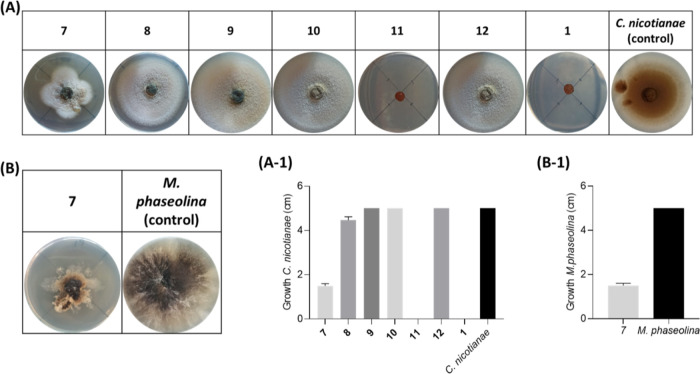
Antifungal Bioassay. (A) and (B) Representative photographs of the antifungal assay against C. nicotianae and M. phaseolina, respectively. Detection of the inhibition of growth of C. nicotianae (cm) and M. phaseolina (cm) (A-1 and B-1, respectively). 7, pinolidoxin; 8, O,O′-diacetylderivate; 9, epi-pinolidoxin; 10, stagonolide C; 11, modiolide A; 12, stagonolide H; and 1, truncatenolide. All compounds were tested at a final concentration of 2.5 × 10⁻³ mol/L. The experiment was performed in triplicate with three independent trials. Data are presented as means ± the standard deviation (n = 4) compared to the control with a p-value < 0.001.

