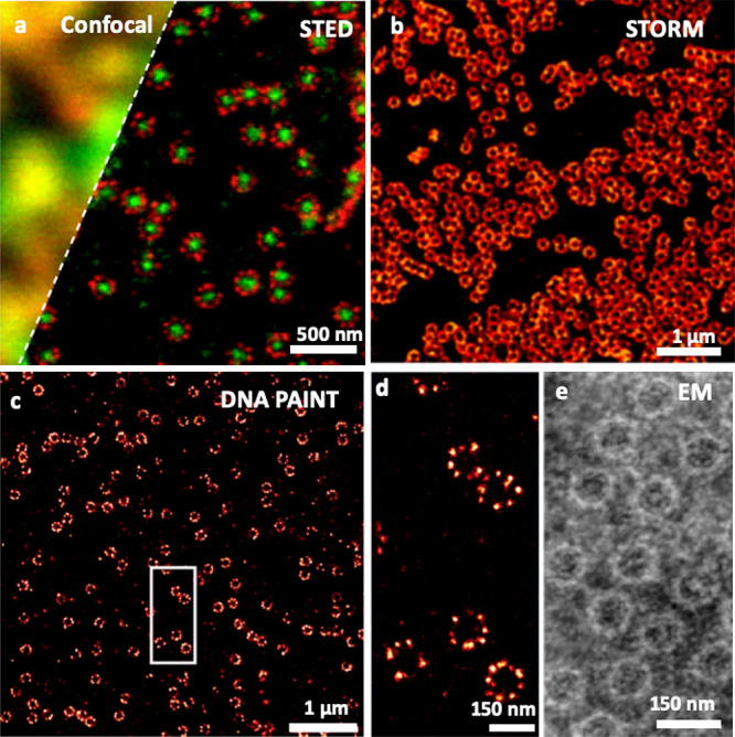
Figure 1.

Super-resolution fluorescence microscopy of nuclear pore complexes with different modalities: STED microscopy (a), compared with confocal microscopy; STORM microscopy (b); DNA PAINT microscopy (c, d). Comparison to electron microscopy using negative staining. Republished with permission of Company of Biologist Ltd. from ref (22). Copyright 2012 The Company of Biologists, permission conveyed through Copyright Clearance Center, Inc.; Adapted with permission from ref (23). Copyright 2019 John Wiley and Sons, https://creativecommons.org/licenses/by/4.0/; Reprinted with permission from ref (24). Copyright 2013 Elsevier.
