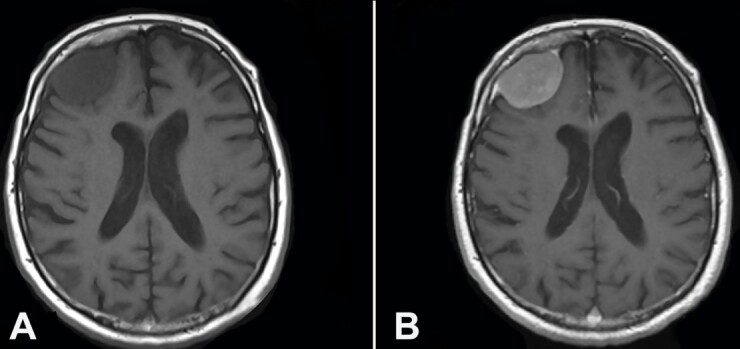
Figure 1. A– Preoperative Axial Brain MRI T1 weighted images showing a frontal hypointense extra-axial right lesion; B – Preoperative Axial Brain MRI T1 weighted with contrast enhancement showing homogeneous enhancement after gadolinium administration of the frontal right lesion with the characteristic dural tail.

