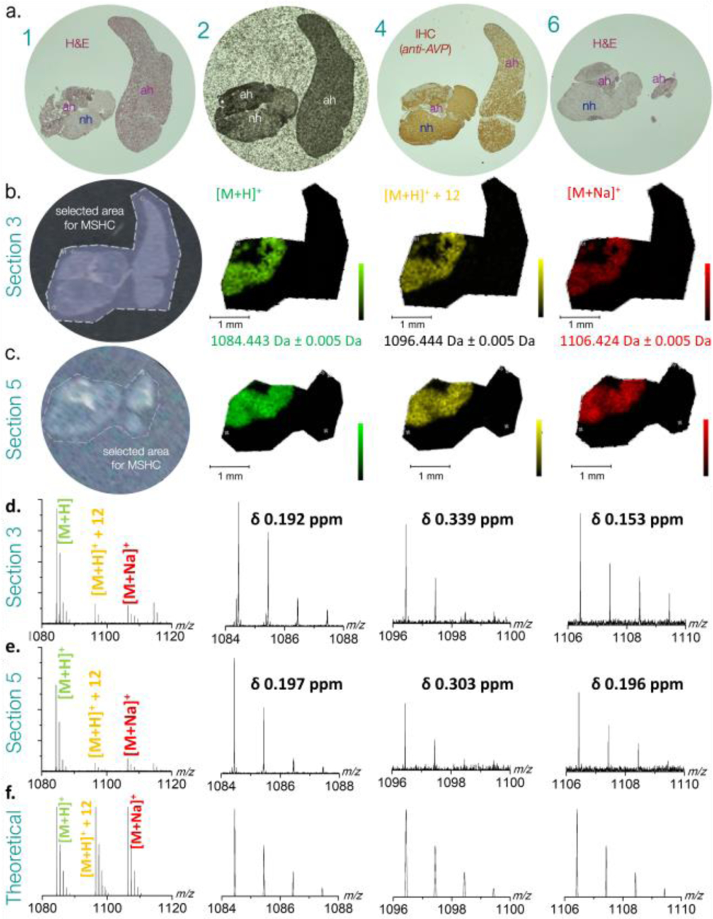
Figure 2.

a. Bright field optical images of human pituitary biopsy tissue sections adjacent to MSHC imaged section 3 and 5. (1) haematoxylin-eosin staining of section 1; (2) unstained section 2 coated with DHB MALDI matrix; (4) immunohistochemically stained section 4 (in between MHSC imaged sections 3 and 5; (6) haematoxylin-eosin staining of section 6, at end of series. b. and c. MSHC experiments showing software selected areas analyzed (left panel) and the respective MALDI-FT ICR MSHC images of protonated (green), Shiff base (yellow) and sodiated (red) ions of Arg-vasopressin ions. Legend: ah, adenohypophysary tumour tissue; H&E, hematoxylin eosin; IHC, immunohistochemistry (using anti-vasopressin polyclonal antiserum). d. and e. showcase mass spectra and experimental isotopic patterns for each species in section 3 and section 5, respectively. f. theoretical isotopic patterns for each species.
