Figure 3.

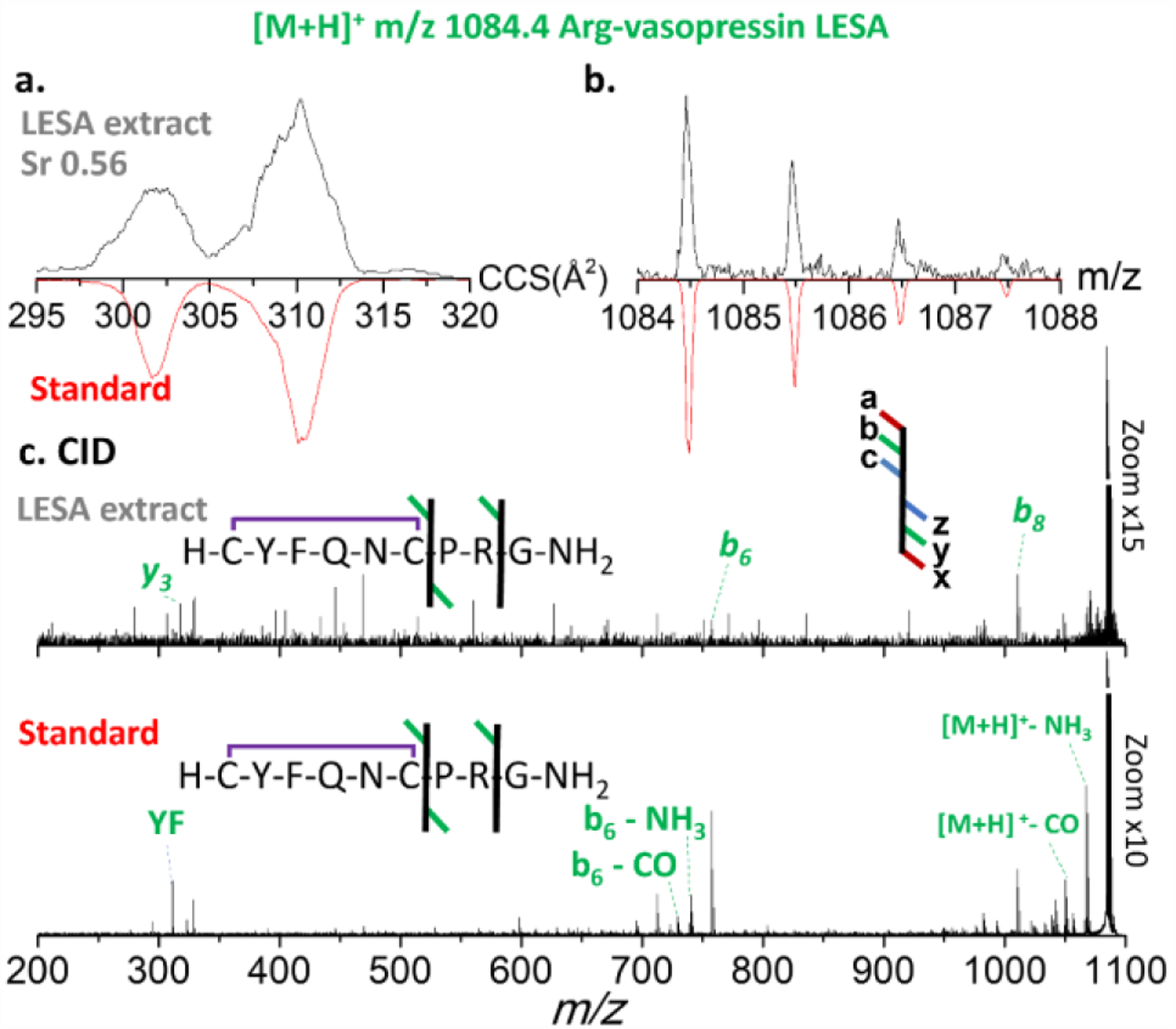
Figure 3. Ion mobility, isotopic and fragmentation patterns of LESA extracted [M+H]+ ions of Arg-vasopressin species. a. ion mobilogram of experimental LESA extracted tissue sample [upper panel, black traces] and of synthetic peptide standard [red traces]; b. isotope pattern of experimental LESA extracted tissue sample [upper panel, black traces] and of synthetic peptide standard [red traces]; c. CID tandem MS of experimental LESA extracted tissue sample [upper panel] and of synthetic peptide standard [lower panel]).
