Figure 4:
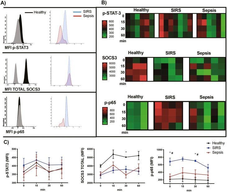
NF-κB activation, but not the canonical STAT3 IL-6 pathway, is increased in patients with SIRS. (A) Histograms showing fluorescence minus one controls (grey) and p-STAT3, SOCS3 and p-p65 (NF-κB) staining in monocytes (FSCmed SSCmed CD45+ CD14+) from healthy volunteers, patients with SIRS and patients with sepsis, after stimulation with IL-6 (100 ng/mL) for 30 min (the histograms represent merged data from all the individuals in each group). (B) Heat maps representing the mean fluorescence intensity (MFI) of p-STAT3, SOCS3, and p-p65 (NF-κB) in monocytes, after activation of peripheral blood with 100 ng/mL of IL-6 for 15, 30, and 60 min (each column represents the results of a healthy volunteer, a patient with SIRS or a patient with sepsis). (C) Activation kinetics of p-STAT-3, SOCS3, and p-p65 (NF-κB) in monocytes, after activation of peripheral blood with 100 ng/mL of IL-6; the graphs represent means and standard error of the mean. Kruskal–Wallis test with Dunn’s multiple comparisons test. For SOCS3: *P < 0.05 in healthy volunteers vs. patients with sepsis at 30 min. For p-p65 (NF-κB): *P < 0.05 in healthy volunteers vs. patients with SIRS at 0 and 30 min. #P < 0.05 in patients with SIRS vs. patients with sepsis at 0 and 30 min.

