Fig. 5.
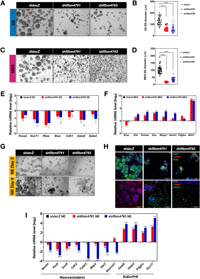
Rbm47 is essential for neuroectoderm and endoderm differentiation of mESCs. A, C and G Phase-contrast images of indicated mESCs directed to DE, MES, and NE lineages. Scale bar − 200 μm. B and D Mean diameter ± S.E.M of the DE-EBs and MES-EBs were calculated using ImageJ software from images acquired from three independent experiments and compared. Unpaired t-test was used to test significance; ns- non-significant; *p < 0.05; **p < 0.01; ***p < 0.001. E, F,and I RT-qPCR profiling of control and shRbm47 mESCs differentiated into indicated lineage. Mean log2 relative expression ± S.E.M values were plotted from three biological replicates. One-way ANOVA followed by Dunnett’s test was used for comparisons; ns- non-significant; *p < 0.05; **p < 0.01; ***p < 0.001; ****p < 0.0001. H Immunostaining of control NE and shRbm47 NE with PAX6, TUBB3, and GATA4 antibodies. Widefield fluorescent images were acquired in the ThermoFisher CellInsight-high content screening platform. Scale bar, 500 μm

