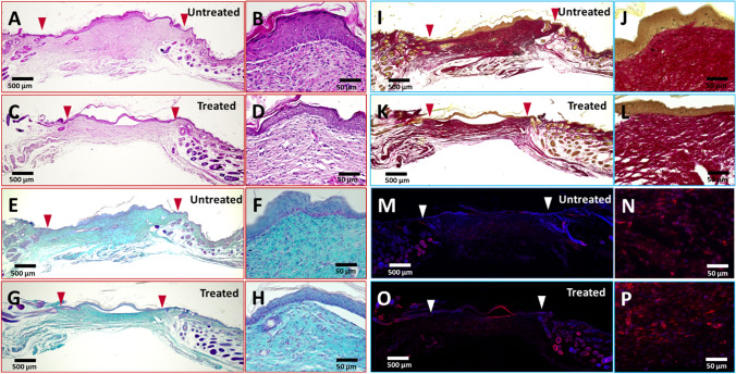
Fig. 6.
Histological staining and immunostaining on moMSCORS untreated and treated groups. Representative photos of histological cross-section of mouse wound bed with and without moMSCORS treatment at the final day 14. Whole range of histological cross-section of the wound skins are shown in (A, C, E, G, I, K, M, O), and higher magnification images of epidermis-dermis interface are shown in (B, D, F, H, J, L, N, P). Arrows demarcate the wound margins. (A, B) H&E staining in moMSCORS untreated group. (C, D) H&E staining in moMSCORS treated group. Nuclei are stained purple blue, and cytoplasm is stained pink. (E, F) Alcian Blue staining in moMSCORS untreated group. (G, H) Alcian Blue staining in moMSCORS treated group. Proteoglycan is stained blue, and nuclei are counterstained pink. (I, J) Picrosirius Red staining in moMSCORS untreated group. (K, L) Picrosirius Red staining in moMSCORS treated group. Collagen filaments are stained bright red, and nuclei are stained black. (M, N) αSMA immunostaining in moMSCORS untreated group. (O, P) αSMA immunostaining in moMSCORS treated group. Arrows demarcate the wound margins. αSMA is labeled with red fluorescence, and the nuclei are counterstained blue

