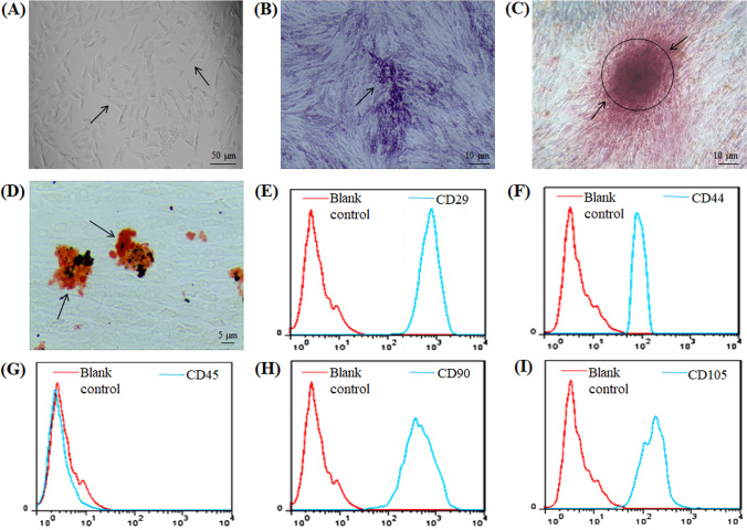
Fig. 1.
Characterization of ADMSCs. A Results of passage 3 ADMSCs observed under light microscope. B The alkaline phosphatase staining of ADMSCs on day 9 of osteogenesis is blue-purple, as shown by arrow. C On day 21 of osteogenic induction, the cells were lamellar with unclear structure, and red calcium nodules were observed in the extracellular matrix stained with Alizarin red S, as shown by arrow. D On day 14 of adipogenesis, fat vacuoles were stained orange by oil red O, as shown by the arrow. E-I Flow cytometry showed positive expression of CD29, CD44, CD90 and CD105, and negative expression of CD45

