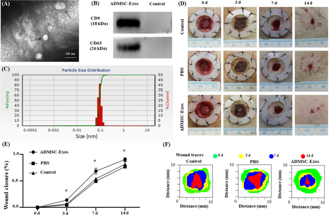
Fig. 2.
Exosomes were successfully isolated from ADMSCs medium; ADMSC-Exos promotes wound healing. A Exosomes were observed by transmission electron microscopy (TEM). B The expression of the exosome protein markers CD9 and CD63 was confirmed by Western blotting. C Nanoparticle tracking analysis (NTA) showed that the ADMSC-Exos distribution diameter ranged from 40 to 100 nm. D, E As can be seen from the wound images at different time points, the wound shrinkage rate of the AMDMSC-Exos group was significantly faster than that of the PBS group and the blank control group. At the 14th day, the remaining wound area was significantly reduced compared with the other two groups. F Traces of wound-bed closure during 14 days in vivo for each treatment category. The data were statistically analyzed using GraphPad. Results are presented as mean ± SD; n = 3; *p < 0.05, compared with PBS and blank control group

