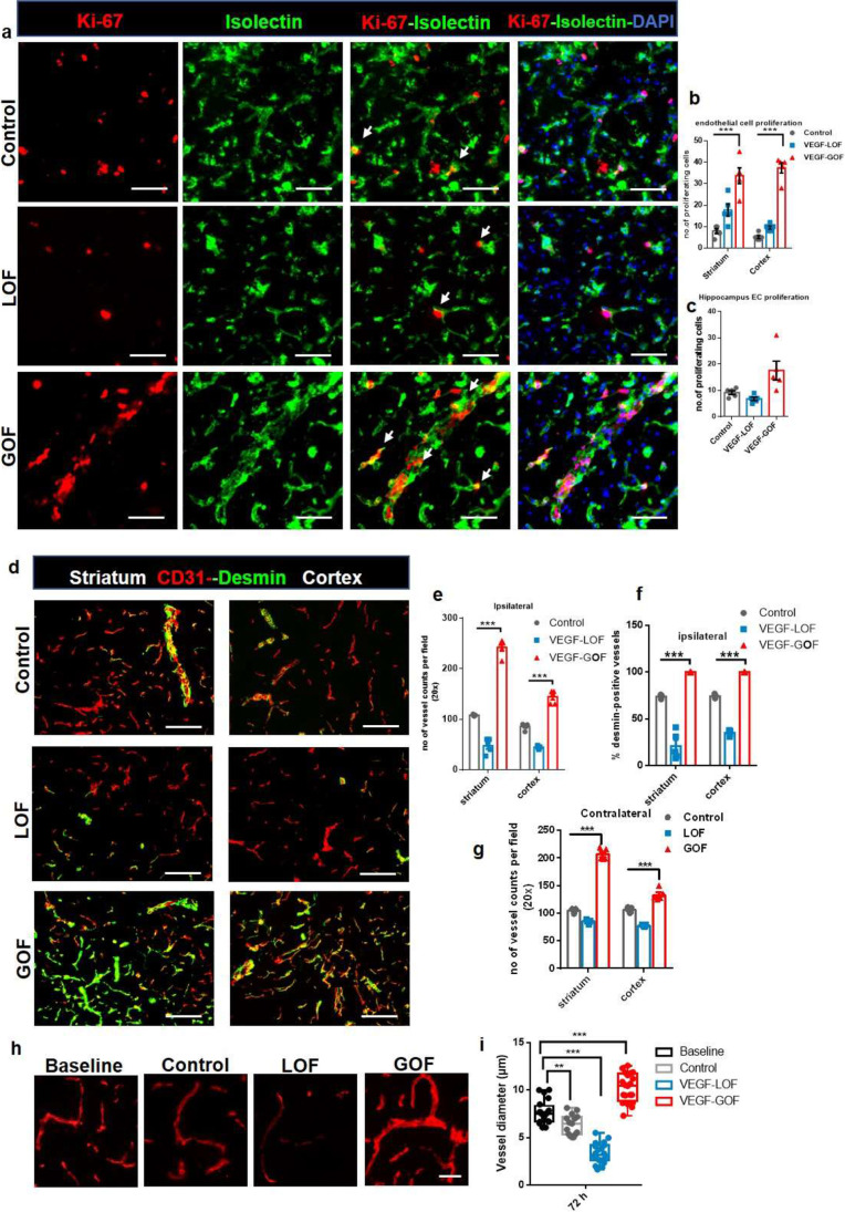
Fig. 2.
VEGF overexpression accelerated vascular recovery via enhanced endothelial–pericyte interactions and blood vessel sealing. (a–c) VEGF overexpression enhances endothelial cell (EC) proliferation. KI-67 labelled cells co-stained with neovascular marker isolectin, showed increased EC proliferation in the VEGF-GOF 72 h post-stroke. On the contrary, VEGF-LOF and control showed fewer proliferating endothelial cells in striatum, cortex, and hippocampus (***P < 0.001). (d) CD31/ desmin co-staining on cryofixed sections shows the enhanced pericyte coverage of cerebral vessel in VEGF-GOF animals compared with LOF and control animals. (e, f) Total number of vessels were increased in the animals with the VEGF overexpression and quantification of desmin-positive vessels show increase in percentage of stable vessels, that is, desmin-covered vessels in the VEGF-GOF (***P < 0.001). (g) VEGF overexpression (GOF) for 28 days lead to an increase in the number of blood vessel in the contralateral hemisphere prior to stroke (***P < 0.001). n = 10 animals per experimental groups. P values were determined by ANOVA. Values are represented as ± SEM. Magnification 200 × ; scale bar 50 μm. (h) Fluorescent images of vessels stained with CD31 showing the vessel diameter in baseline, control, VEGF-LOF and VEGF-GOF animals, scale bar 15 µm. (i) The box and whiskers plot show the blood vessel diameter in each experimental group (**P < 0.01, ***P < 0.001), P values were determined by one-way ANOVA

