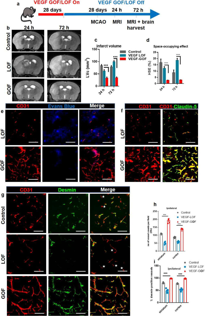
Fig. 5.
VEGF-induced vascular stability is not reversible after VEGF withdrawal (VEGF on > off). (a) Schematic timeline representation of the experimental procedure. Transgenic expression of VEGF or I-sVEGF-R1 was switched on for 28 days, and then turned off for 28 days before inducing 60 min MCAO. (b) T2-weighted magnetic resonance images (MRI), representative MRI from control, VEGF-LOF (on > off) and VEGF-GOF (on > off) animal 24 h and 72 h post-stroke. (c, d) quantitation of the lesion volume corrected (LVc, mm3) and space-occupying effect attributable to edema (% HSE) after stroke. Infarct size is significantly reduced in VEGF-GOF (on > off) already from 24 h post-stroke, whereas VEGF-LOF (on > off) show a significant increase in the infarct volume 24 h post-stroke and the infarct size further increase 72 h post-stroke. The space-occupying effect in VEGF-GOF (on > off) is significantly reduced 24 h post-stroke indicating a decrease in the volume of brain swelling, on the other hand blocking VEGF signaling (LOF on > off) aggravated brain swelling. n = 5 animals per experimental groups. P values were determined by ANOVA (***P < 0.001). Values are represented as ± SEM. (e) Confocal analysis of Evans Blue and CD31 showing an increased in cerebrovascular permeability in the ischemic core in VEGF-LOF (on > off) versus VEGF-GOF (on > off). n = 5 in both experimental groups. (f) Confocal images of CD31-claudin-5 showed an increase in the tight junction protein claudin-5 expression in VEGF-GOF (on > off), whereas blood vessels in VEGF-LOF (on > off) have lower claudin-5 coverage 72 h post-stroke. (g) Confocal images show an increase in pericyte coverage of cerebral vessel in VEGF-GOF (on > off) compared with VEGF-LOF (on > off) and control. Star shape marks the microvessels without the pericyte coverage. (h, i) Total number of vessels were increased in the VEGF-GOF (on > off animals) and quantification of desmin-positive vessels show that all the blood vessels are desmin-covered. VEGF-LOF (on > off) showed significant decrease in the number of vessels and the desmin-positive vessels were significantly decreases 72 h when compared to the control and VEGF-GOF (on > off). n = 5 animals per experimental groups. P values were determined by ANOVA (***P < 0.001). Values are represented as ± SEM. Scale bar 20 μm

