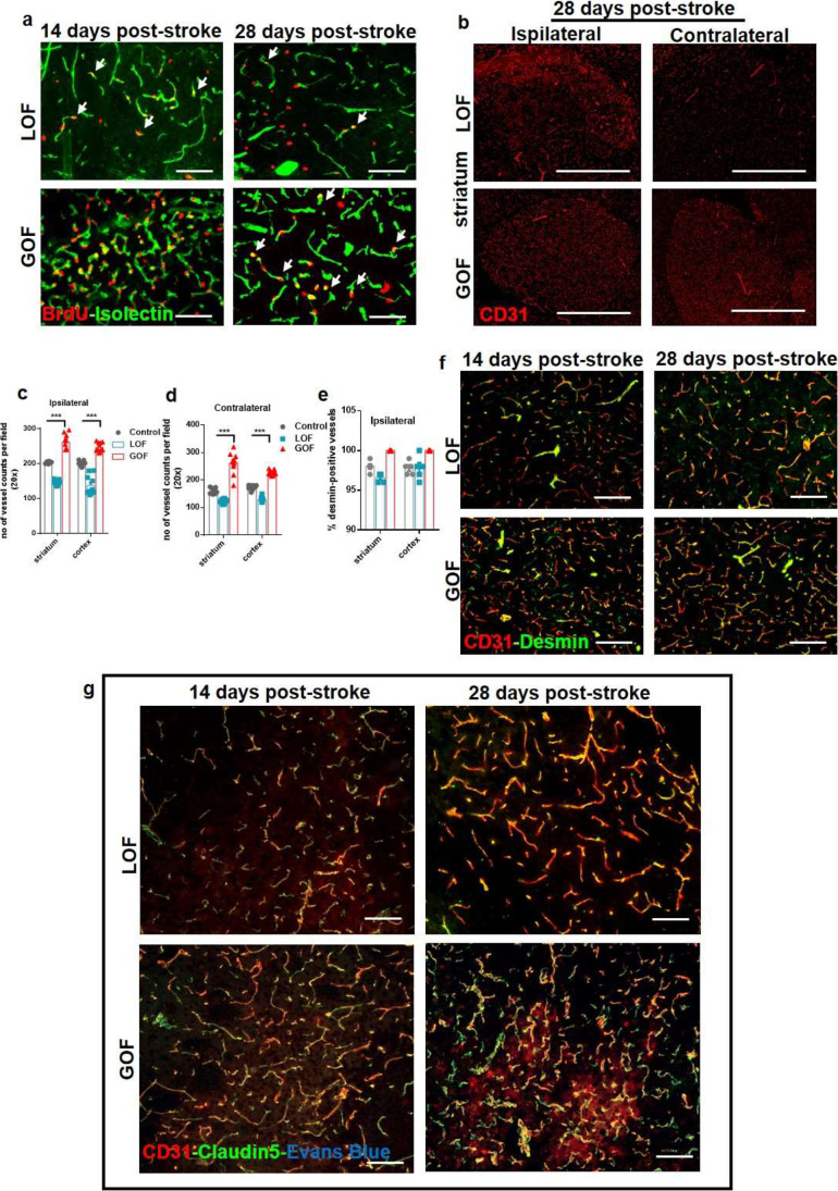
Fig. 7.
VEGF-induced long-term changes in the late phase of stroke. (a) BrdU labelled cells co-stained with neovascular marker isolectin, showed increased EC proliferation in the VEGF-GOF animals 14 days and 28 days post-stroke. VEGF-LOF animals showed fewer EC proliferation on day 14 and 28 after stroke. (b-d) CD31 staining and quantification data show an increase in the number of blood vessels in the both ipsilateral and contralateral hemisphere of the VEGF-GOF 28 days post-stroke, whereas VEGF-LOF show post-stroke neovascularization only in the ipsilateral region (Scale bar 100 μm). P values were determined by ANOVA (***P < 0.001). Values are represented as ± SEM. (e, f) CD31/desmin co-staining on cryofixed sections and quantification of desmin-positive vessels show no difference in percentage of stable vessels in VEGF-GOF, VEGF-LOF and control. n = 10 animals per experimental groups. P values were determined by ANOVA. Values are represented as ± SEM. (g) CD31/claudin-5 co-staining on cryofixed sections showed no difference in tight junction protein coverage in VEGF-GOF and VEGF-LOF 14- and 28-day post-stroke. No Evans Blue extravasation was observed at these timepoints. Scale bar 50 μm

