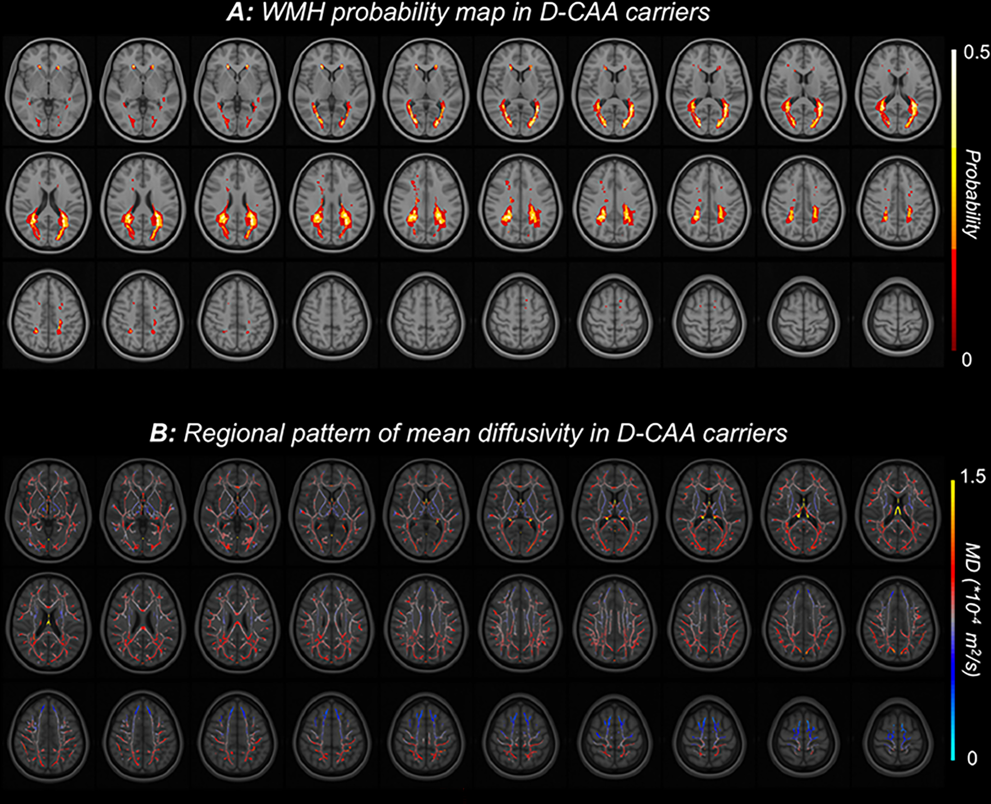
Figure 1:

Spatial patterns of white matter injury in D-CAA. A: FLAIR-based white matter hyperintensity (WMH) probability map in D-CAA carriers showing a posterior predominance. Individual images were placed in MNI space, and voxels are colored by the probability of a white matter lesion being present in this cohort. B: Diffusion based mean diffusivity in the white matter skeleton. Individual mean diffusivity images were placed in MNI space, and voxels are colored by the average value across D-CAA carriers in this cohort.
