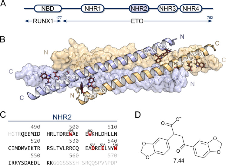
Figure 1.
Schematic representation of RUNX1/ETO tetramerization. (A) Depiction of the chimeric RUNX1/ETO fusion protein containing the nucleotide-binding domain (NBD) and four nervy homology regions (NHR). The NHR2 domain (marked in blue) mediates homo-tetramerization. (B) X-ray crystal structure (PDB ID 1WQ6) of the NHR2 domain. For the clarity of the tetrameric α-helical bundle, one antiparallel α helical pair is shown in ribbon view and the other in surface view. The five predicted hot spot residue11 side chains (W498, W502, D533, E536, and W540) are shown in stick representation and colored by elements (C) Primary amino acid sequence of the NHR2 domain shown in black, hot spot residues are colored in red, other ETO residues in gray. (D) Molecular structure of 7.44.

