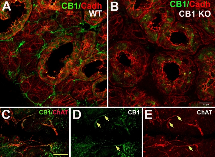
Figure 2.
CB1 protein is expressed in a subset of cholinergic axons in submandibular gland. (A,B) Left panel shows CB1 (green) and cadherin (red) in wild type (WT) mouse submandibular gland. Right panel shows that CB1 staining in axon-like processes is absent submandibular gland from CB1 knockout mouse. (C) Double staining in submandibular gland of the mouse shows expression of CB1 (green) and choline acetyl transferase (ChAT, red), a marker for cholinergic axonal inputs. (D,E) CB1 (green) staining is seen in processes of ChAT-positive axons (arrows). Scale bar: (A,B) 25 µm; (C–E) 40 µm. Images processed using Adobe Photoshop vsn. 21.2 and FIJI (vsn 2.3.0/1.53q, available at https://imagej.net/Fiji/downloads).

