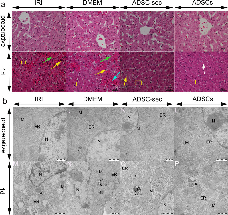
Fig. 1.
Histopathological changes and ultrastructural changes in the liver post-IRI. a: HE-stained liver tissues. Green arrows indicate necrosis, the white arrow indicates hepatocyte vacuolar degeneration, the blue arrow indicates hemorrhage, yellow rectangle indicates hepatocyte swelling, and yellow arrows indicate inflammatory cell infiltration (Magnification × 400). b: Transmission electron microscopy micrographs of the liver. A: Autophagy structure; N: nucleus; ER: endoplasmic reticulum; and M: Mitochondria (magnification: 12,000 ×). IRI: ischemia–reperfusion injury; ADSC-sec: adipose-derived mesenchymal stromal cell-secretome; and ADSCs: adipose-derived mesenchymal stromal cells

