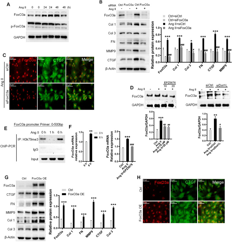
Fig. 7.
Dot1L directly binding to the promoter regions of FoxO3a promotes ECM deposition. A FoxO3a is upregulated in NRCFs treated with 2 μmol/L Ang II for 24 and 48 h. FoxO3a and its phosphorylation form were analyzed by western blot. B, C Knockdown of FoxO3a attenuates ECM deposition in Ang II-treated NRCFs. FoxO3a siRNA and control siRNA were transfected into NRCFs, followed by Ang II stimulation for 48 h. The protein expressions of FoxO3a, Col 1, Col 3, FN, MMP9 and CTGF were analyzed by western blot (B); Representative images of immunofluorescence staining of FoxO3a and CTGF in Ang II-stimulated NRCF (C). D NRCFs were pretreated with EPZ5676 (10, 20 μmol/L) for 4 h or transfected with siDot1L for 12 h, then incubated with 2 μmol/L Ang II for 48 h. FoxO3a was analyzed by western blot. E ChIP-PCR showing H3K79me3 gain in promoter of FoxO3a in Ang II-treated NRCFs. NRCFs treated with Ang II for 1 and 6 h were used for ChIP assay with antibody against H3K79me3. F mRNA level of FoxO3a was quantified by qRT-PCR after inhibition of Dot1L with EPZ5676. Above all data are represented as means ± SD. ANOVA, *p < 0.05, **p < 0.01, ***p < 0.001 versus Ctrl or Ctrl + siCtrl, #p < 0.05, ##p < 0.01, ###p < 0.001 versus Ang II or Ang II + siCtrl, each acquired from three individual experiments. G, H Overexpression of FoxO3a promotes fibrosis response in NRCFs. NRCFs were transfected with lentivirus containing FoxO3a overexpression plasmid for 24 h, and then cultured with completed medium for 48 h. The protein expressions of FoxO3a, CTGF, FN, MMP9, Col 1 and Col 3 were analyzed by western blot. Data are represented as means ± SD. Student’s t-test, ***p < 0.001 versus Ctrl, each acquired from three individual experiments (G); immunofluorescence double staining of FoxO3a and CTGF (H)

