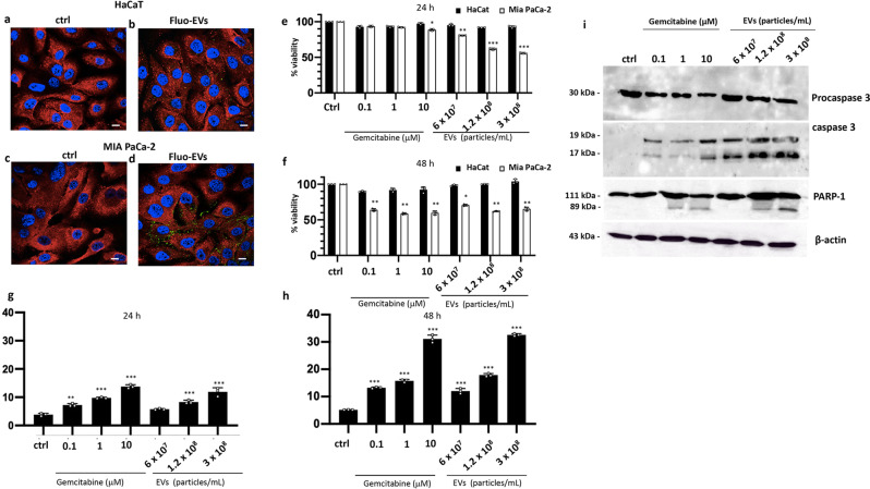
Fig. 3. Uptake and biological effects of HR-derived EVs in human control and cancer cell lines.
a–d Confocal analysis of HaCaT and MIA PaCa-2 cells treated with Bodipy-stained EVs (Fluo-EVs) (1.2 × 10⁸ particles/mL) for 24 h. Cells have been stained for annexin A1 protein in red. Nuclei were stained with DAPI. Magnification 63×/1.4 numerical aperture. Scale bar = 100 μm. e, f MTT colorimetric assay on HaCaT and MIA PaCa-2 cells after 24 h and 48 h of EV treatments, respectively. Absorbance relative to controls was used to determine the percentage of cells treated with varying concentrations of gemcitabine and EVs. g, h Analyses of apoptotic cells by cytofluorimetric assay in gemcitabine- and EVs-treated MIA PaCa-2 cells upon 24 h and 48 h exposure. The values reported in the graphs are the mean ± SD from at least 3 independent experiments performed in technical triplicates. The asterisks denote significant differences between treatments and untreated controls (*p < 0.05; **p < 0.01; ***p < 0.001) according to Student’s t-test. i Western blot analyses of protein extracts from cells treated for 24 h with different concentrations of gemcitabine and EVs. Levels of cleaved proteins involved in the apoptosis (Procaspase 3 and PARP-1) were evaluated. β−actin was used to check equal loading of protein extracts.

