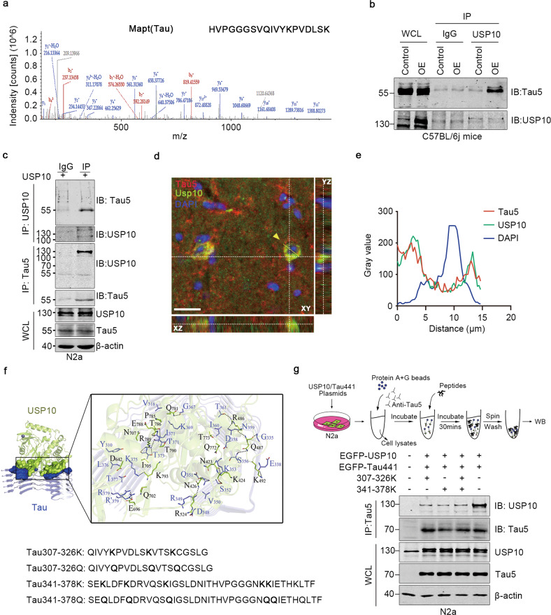
Fig. 5. Synthesized peptides block USP10 interaction with Tau.
a Mass spectrometry analysis of USP10-interacting proteins. The detected Mapt (Tau) typical mass spectrometry peptide spectra were listed. The list of USP10-interacting proteins is provided in Supplementary Table S3. b USP10 interacts with Tau5 in C57BL/6j mice tissue. AAV9-USP10 was injected into the hippocampus of wild-type C57BL/6j mice, tissues were immunoprecipitated with USP10 and subjected to western blotting. c USP10 interacts with Tau5 in N2a cells. Immunoblots of co-immunoprecipitation (upper) or reverse co-immunoprecipitation (lower) of USP10 and Tau5. WCL were analyzed using western blotting. d Confocal microscopy Z-stack imaging to hippocampus region of 13-month-old APP/PS1 mice. Immunostaining of Tau5 (Red), USP10 (Green), and DAPI (blue). X–Z, X-Y and Y-Z directions were shown. Yellow dotted lines represent the Z-depth of the 5 µm optical slice, and the yellow line represents the statistical area. The scale bar is 20 μm. e Statistical analysis of USP10, Tau5, and DAPI immunoreactivity. f Visualization of molecular docking of USP10 and Tau5 of the lowest energy, and the design of a series of blocking peptides targeting the USP10-Tau interaction interface. g Upper panel was a schematic of experimental process. N2a cells were co-transfected with USP10 and Tau. Cytoplasmic extracts were immunoprecipitated with a Tau5 antibody. The peptides Tau 307-326 K, Tau341-378K, or Tau 307-326 K + Tau341-378K at a concentration of 1 mM for 30 min at room temperature were used to compete with the USP10-Tau interaction in vitro. IP and WCL samples were blotted with the indicated antibodies.

