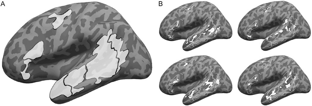
Fig. 1.
Defining participant-specific fROIs in the core language network. All images show approximate projections from functional volumes onto the surface of an inflated average brain in common (MNI) space. (A) Group-based masks used to constrain the location of fROIs. These masks were derived from a probabilistic group-level representation of the sentences > nonwords localizer contrast in a separate sample, following (Fedorenko et al., 2010). Contours of these masks are depicted in white in B. (B) Example fROIs of four participants. Note that, because data were analyzed in volume (not surface) form, some parts of a given fROI that appear discontinuous in the figure (e.g., separated by a sulcus) are contiguous in volumetric space.

