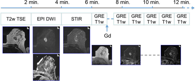
Fig. 1.
A 15-min clinical protocol for breast magnetic resonance imaging (MRI). All predictive/prognostic breast MRI information demonstrated in the next figures can be derived from a one-stop shop clinical protocol as shown in this figure. The protocol starts with an unenhanced T2-weighted turbo spin-echo sequence (T2w TSE). Diffusion-weighted imaging (DWI) and short-tau inversion recovery (STIR) are optional but highly recommend. On T2-weighted images, a mass lesion is diagnosed, with perifocal oedema. Next, contrast-enhanced dynamic scanning is performed using a T1-weighed gradient-echo (GRE) sequence before/after the intravenous administration of 0.1 mmol/kg of a Gd-based contrast agent. There is evidence of washout, perifocal oedema, and central necrosis (rim sign). The last two descriptors are imaging biomarkers associated with increased probability of high-grade and nodal-positive invasive cancers. Washout is a strong predictor of poor outcome and is associated with a higher likelihood of metachronous metastasis (see also Figs. 5 and 6). Example taken from ref [2], with permission (Dietzel et al. Insights Imaging 2018)

